
Figures & data
Figure 1. Conditioned medium from TAMs upregulates the expression of Gli2 and IGF-II and promotes migration and invasion of Huh-7 cells
(A-B) THP-1 cells were differentiated and polarized into M2 macrophages, and the expression levels of CD68 and CD206 were detected by flow cytometry (A) and quantified (B). (C) After differentiation and polarization, the medium of THP-1 cells was collected, and the concentrations of IL-10 and TGF-β1 were measured by ELISA. (D-E) After treatment with conditioned medium of TAMs (TAMs-CM), the Huh-7 cells were collected, and the levels of TGF-β1, Gli2 and IGF-II were examined by qPCR (D) and western blotting (E). (F) The migration and invasion abilities of Huh-7 cells treated with TAMs-CM were detected by Transwell assay. (G) The migration of Huh-7 cells treated with TAMs-CM was detected by scratch assay. (H) The proliferation of Huh-7 cells treated with TAMs-CM was detected by CCK-8 assay. *** p < .001, ** p < .01, * p < .05.
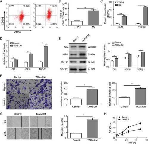
Figure 2. TGF-β1 signaling mediates the effect of TAMs-CM on Gli2 expression and Huh-7 cell migration and invasion
Huh-7 cells were treated with the conditioned medium of TAMs and with different concentrations of the TGF-β1 inhibitor LY364947. (A) The level of Gli2 mRNA was measured by qPCR. (B) The levels of Gli2 and TGF-β1 protein were measured by western blotting. (C-D) The invasion and migration abilities were measured by Transwell assays; the quantification is shown in (D). (E-F) The migration of Huh-7 cells was measured by scratch assay; the quantification is shown in (F). *** p < .001, ** p < .01, * p < .05.
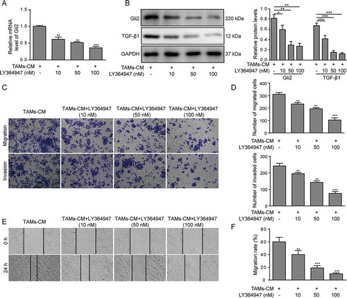
Figure 3. Gli2 induces IGF-II expression to promote Huh-7 cell migration and invasion
(A-C) Huh-7 cells were transfected with sh-Gli2, and the levels of Gli2 and IGF-II mRNA were measured by qPCR (A-B); the levels of Gli2 and IGF-II protein were measured by western blotting (C). (D-E) The migration and invasion abilities of Huh-7 cells were measured by Transwell assay. (F-G) The migration of Huh-7 cells was confirmed by scratch assay. (H) The protein levels of E-cadherin, vimentin and MMP-9 were detected by western blotting. *** p < .001, ** p < .01, * p < .05.
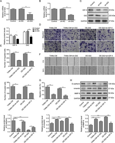
Figure 4. IGF-II promotes migration and invasion of Huh-7 cells via the ERK1/2 signaling pathway
(A) Huh-7 cells were treated with IGF-II or its vehicle, and the expression and phosphorylation levels of ERK1/2 were measured by western blotting. (B-E) Huh-7 cells were treated with IGF-II or the ERK1/2 inhibitor PD98059 or with both IGF-II and PD98059. The invasion and migration of Huh-7 cells were measured by Transwell assay (B, C), and the migration was measured by scratch assay (D, E). (F) The activation of ERK1/2 and the expression of E-cadherin, vimentin and MMP-9 were analyzed by western blotting. *** p < .001, ** p < .01, * p < .05.
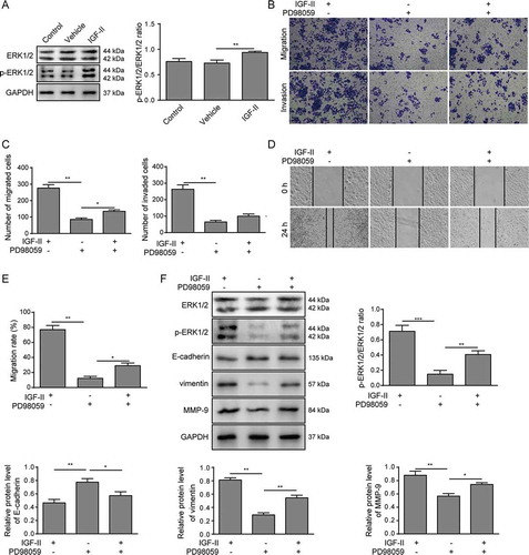
Figure 5. TAMs-CM promotes the migration and invasion of Huh-7 cells through the Gli2/IGF-II/ERK1/2 axis
(A-B) Huh-7 cells were treated with TAMs-CM, TAMs-CM and PD98059, Gli2 overexpression, or Gli2 overexpression and PD98059. Cell invasion and migration were measured by Transwell assay. (C-D) Cell migration was confirmed by scratch assay. (E) The activation of ERK1/2 and the expression of E-cadherin, vimentin and MMP-9 were analyzed by western blotting. *** p < .001, ** p < .01, * p < .05.
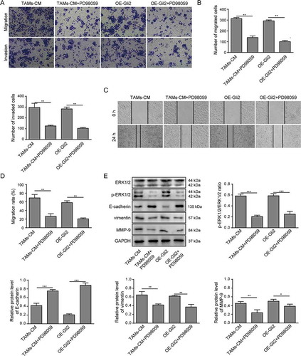
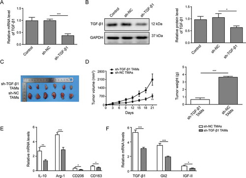
Figure 6. TAMs promote Huh-7 tumor growth in vivo mediated by TGF-β1
(A-B) TAMs were infected with lentivirus harboring scrambled control shRNA (sh-NC) or shRNA targeting TGF-β1 (sh-TGF-β1). The expression of TGF-β1 was examined by qPCR (A) and western blotting (B). (C) An image of the tumors collected after the mice were sacrificed. (D) The growth rate and weight of tumors in each group. (E) The expression levels of M2 macrophage markers IL-10, Arg-1, CD206 and CD163 in isolated tumors, as detected by qPCR. (F) The expression levels of TGF-β1, Gli2 and IGF-II in tumors detected by qPCR. *** p < .001, ** p < .01, * p < .05.
Supplemental material