Figures & data
Figure 1. An aCGH-based analysis of DNA copy-number alteration frequencies in 50 endometrial carcinoma samples. Gains or losses for each measured sequence are shown on the y-axis, with sequences being aligned along the x-axis in chromosomal order. The significance threshold is represented by a dashed line. Significant DNA copy-number gain frequencies are marked by red lines, while significant DNA copy-number losses are marked by green lines. Non-significant changes are marked in gray. Vertical bars demarcate different chromosomes, with dashed vertical bars marking the separation between short and long arms of individual chromosomes
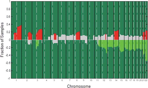
Table 1. Gene alterations in endometrial carcinoma
Table 2. SOX8 expression in NE, HE, AHE, and EC tissues
Table 3. Correlation between SOX8 expression and clinicopathological parameters in EC cases
Figure 2. SOX8 expression levels in human EC tissue samples correlate with aggressive clinicopathological findings. (a) An IHC approach was used to measure SOX8 levels in TMAs. The resultant data revealed that SOX8 levels were higher in EC and AHE samples relative to HE and NE samples. (b) The expression of SOX8 in EC samples was significantly higher than that in NE samples. (c) SOX8 overexpression was linked to high EC histological grade. Left: Representative images of EC tissue SOX8 staining (G1 – G3). Right: Elevated SOX8 expression was confirmed to be significantly associated with high EC histological grade in these patient samples. (d) SOX8 overexpression was associated with EC lymph node metastasis. Left: Representative SOX8 staining in LN− and LN+ EC samples. Right: Elevated SOX8 expression was confirmed to be significantly associated with EC lymph node metastasis in these patient samples. (e) The relationship between SOX8 expression and post-operative EC patient OS was assessed using a Kaplan-Meier curve. (**p< .01, *p< .05)
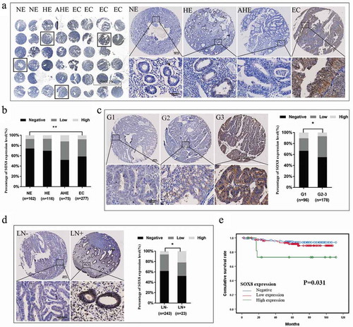
Figure 3. SOX8 controls EC cell proliferation and growth. (a) Endogenous SOX8 levels in ECC-1, Ishikawa, HEC1A, HEC1B, and KLE cells were assessed by Western blotting. SOX8 knockdown in HEC-1B cells (b,d) and SOX8 overexpression in Ishikawa cells (c,e) was confirmed via Western blotting and qPCR. A CCK8 assay revealed that SOX8 knockdown significantly impaired cell proliferation (f), whereas SOX8 overexpression had the opposite effect (g). A colony-formation assay indicated that SOX8 knockdown impaired EC cell growth (h), while SOX8 overexpression significantly enhanced proliferation (i). Data are means ± SD from triplicate experiments. #p < .05 vs. control; *p < .05 vs. NC control
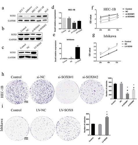
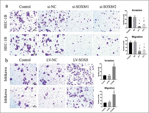
Figure 4. SOX8 drives EC cell migratory and invasive activity in vitro. (a) The invasive and migratory activity of HEC-1B cells was significantly decreased following SOX8 knockdown. (b) Overexpression of SOX8 significantly enhanced Ishikawa cell migratory and invasive activity (×100)