
Figures & data
Figure 1. Colorectal cell lines are efficiently transduced and express the transgenes
HCT116wt, HCT116−/- and HT29 (p53 R273H) cells were seeded in 24 wells plate (5.104 cells per well). After 24 h, cell number was verified and then transduced using AdRGD-PG-eGFP (G), AdRGD-PG-p53 + AdRGD-PG-eGFP (P), AdRGD-PG-hIFNβ + AdRGD-PG-eGFP (I) AdRGD-PG-p53 + AdRGD-PG-hIFNβ (C) or no vector (O). Cells and supernatant were harvested 48 h after transduction and analyzed by the respective methodology. A. Western blot showing p53 expression. B. IFN-β measurement in cell supernatant via ELISA. These data are qualitative and included to confirm expression, thus do not conclusively indicate relative expression levels. C. Percentage of positive cells for GFP detected by flow cytometry. D. Intensity of GFP fluorescence (AU, arbitrary units). * p < .05 One Way Anova; Tukey posttest. In panels C and D, results represent the average and standard deviation of three independent experiments.
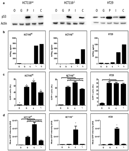
Figure 2. Cellular response to transgene activity
HCT116wt, HCT116−/- and HT29 (p53 R273H) cells were seeded in 24 wells plate (5.104 cells per well). After 24 h, cell number was verified and then transduced using AdRGD-CMV-LacZ (L), AdRGD-PG-eGFP (G), AdRGD-PG-p53 + AdRGD-PG-eGFP (P), AdRGD-PG-hIFNβ + AdRGD-PG-eGFP (I) or the combination AdRGD-PG-p53 + AdRGD-PG-hIFNβ (C). Cells were harvested at indicated times and analyzed using the respective methodology. A. Viable cells counted 72 h post-transduction. B. Number of colonies formed. C. Viability is measured via MTT 96 h post-transduction. * p < .05 One Way Anova; Tukey posttest. Results represent the average and standard deviation of three independent experiments.
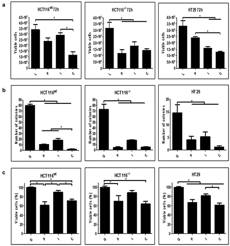
Figure 3. Induction of cell death markers upon treatment with adenovirus
HCT116wt, HCT116−/- and HT29 (p53 R273H) cells were seeded in 24 wells plate (5.104 cells per well). After 24 h, cell number was verified and then transduced using AdRGD-CMV-LacZ (L), AdRGD-PG-eGFP (G), AdRGD-PG-p53 + AdRGD-PG-eGFP (P), AdRGD-PG-hIFNβ + AdRGD-PG-eGFP (I) or the combination AdRGD-PG-p53 + AdRGD-PG-hIFNβ (C). Cells were harvested at the indicated times and analyzed using the respective methodology. A. Percentage of sub G1 (hypodiploid) cells was detected by PI staining and flow cytometry 72 h post-transduction. B. Percentage of cells positive for Annexin V staining and negative for PI staining at 72 h as detected by flow cytometry. C. Percentage of cells positive for active caspase 3 and 7. * p < .05 One Way Anova; Tukey posttest. Results represent the average and standard deviation of three independent experiments.
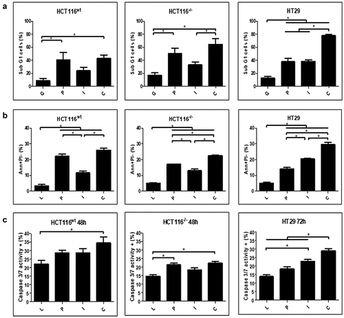
Figure 4. Emission of immunogenic cell death markers in response do gene transfer
HCT116wt, HCT116−/- and HT29 (p53 R273H) cells were seeded in 24 wells plate (5.104 cells per well). After 24 h, cell number was verified and then transduced using AdRGD-PG-Luc (L), AdRGD-PG-p53 + AdRGD-PG-eGFP (P), AdRGD-PG-hIFNβ + AdRGD-PG-eGFP (I) AdRGD-PG-p53 + AdRGD-PG-hIFNβ (C) or no vector (O). Cells and supernatant were harvested at 72 hours post transduction and analyzed using the respective methodology. A. Percentage of cells with calreticulin (CRT+) exposure on the cell surface as observed by flow cytometry. (B) Detection of ATP measured as intensity of luciferase activity (AU, arbitrary units) and (C) release of HMGB1 measured by Western blot using equal quantities of conditioned medium. * p < .05 One Way Anova; Tukey posttest. Results represent the average and standard deviation of three independent experiments.
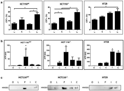
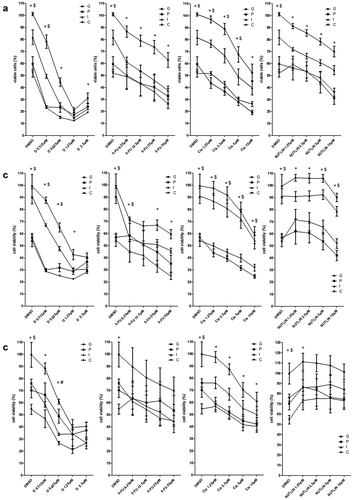
Figure 5. Association of chemotherapy with gene transfer
Cell lines were seeded (5000 cells per well) and transduced using AdRGD-PGeGFP (G) AdRGD-PGp53 + AdRGD-PG-eGFP (P), AdRGD-PG-hIFNβ + AdRGD-PG-eGFP (I) AdRGD-PG-p53 + AdRGD-PG-hIFNβ (C). After 24 hours incubation, doxorrubicin (D) cisplatina (Cis), 5-fluorouracil (5-FU), Nutlin-3 (NUTLIN) or the diluent (DMSO) were added at the concentrations indicated. Then, after 72 hours incubation, MTT assay was performed and viability normalized against the GFP control. (A) HCT116wt, (B) HCT116−/-and (C) HT29. + (C ≠ G); # (C ≠ P); $ (C ≠ I); p < .05 Two Way ANOVA, Bonferroni posttest. Results represent the average and standard deviation of four independent experiments.
Supplemental material