Figures & data
Table 1. Primers used in RT-PCR analysis
Figure 1. Expression of TRIM11 and DUSP6 in the lung tissue of patients with NSCLC, assessed by IHC assay. Ten cases of tumorous and precancerous lung tissues were analyzed. The representative images and the quantification of positive staining are shown. Magnification: ×200. **P < .01 vs. precancerous lung tissue
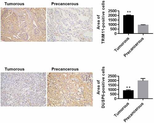
Figure 2. Knockdown of TRIM11 inhibited the tumorigenesis of NSCLC cells, up-regulated DUSP6, and inhibited ERK1/2 activity. (a) mRNA levels of TRIM11, measured by RT-PCR. (b) Proliferation, (c) colony formation, and (d) apoptosis of A549 and H446 cells, assessed by CCK8, colony formation assay, and flow cytometry, respectively. (e and f) siTRIM11 inhibited the glucose metabolism of A549 and H446 cells by decreasing the secretion of LD and the uptake of 2-NBDG. (g) Western blot analysis showed that siTRIM11 remarkably down-regulated TRIM11, PKM2, and ERK1/2, and up-regulated DUSP6 in A549 and H446 cells. **P < .01 vs. siNC

Figure 3. Overexpression of TRIM11 promoted the tumorigenesis of NSCLC cells, down-regulated DUSP6, and promoted ERK1/2 activation. H1975 cells were infected with lentivirus overexpressing TRIM11. (a) RT-PCR showing the mRNA levels of TRIM11. (b) CCK8 for proliferation assay; (c and d) flow cytometry for apoptosis assay; (e) LD secretion, assessed by a commercial Kit; and (f) 2-NBDG uptake, measured by a biochemical analysis method. (g) Western blot analysis showed that TRIM11 remarkably up-regulated TRIM11, PKM2, ERK1/2, and down-regulated DUSP6 in H1975 cells. (h) RT-PCR showing the mRNA levels of TRIM11, PKM2, and DUSP6. ##P < .01 vs. vector
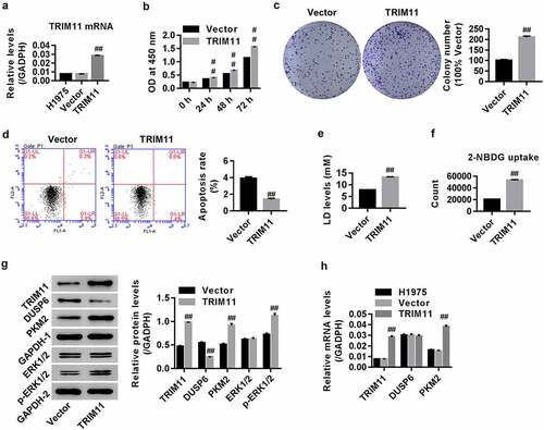
Figure 4. The DUSP6 and ERK1/2 inhibitor dn-MEK1 attenuated TRIM11-induced tumorigenesis of H1975 cells. (a) Successful DUSP6 overexpression in H1975 cells. Both DUSP6 and dn-MEK1 significantly inhibited (b) the secretion of LD, (c) the uptake of 2-NBDG, and (d) the proliferation of H1975, but decreased (e) H1975 apoptosis. (f) Western blot analysis showed that DUSP6 and dn-MEK1 remarkably down-regulated PKM2 and p-ERK1/2 in H1975 cells transfected with TRIM11. ##P < .01 vs. vector, ++P < .01 vs. TRIM11
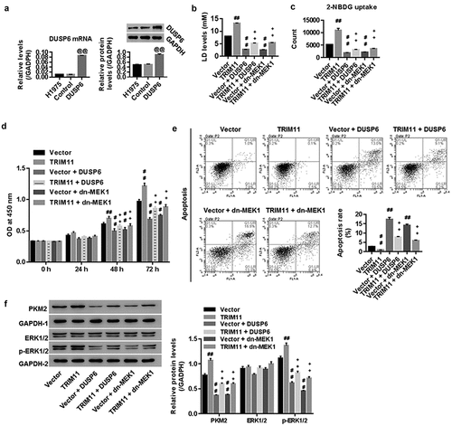
Figure 5. TRIM11 was associated with DUSP6, and regulated the ubiquitinoylation of DUSP6 in human H1975 cells transfected with TRIM11. Following co-immunoprecipitation with (a) anti-DUSP6 antibody and (b) anti-TRIM11 antibody, the presence of TRIM11 and DUSP was measured by western blot. (c) The presence of DUSP6 in TRIM11-transfected HCT116 cells was immunoprecipitated with DUSP6 antibodies and immunoblotted with anti-ubiquitin antibodies
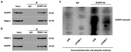
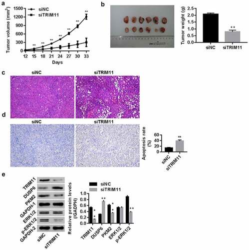
Figure 6. Knockdown of TRIM11 inhibited the tumorigenicity of A549 cells in a xenograft model. Nude mice were injected with siNC/siTRIM11-transfected A549 cells (5 × 10⁶ cells, 100 μl) (n = 6 in each group). (a) Tumor volume (mm3) from the 12th to 33rd day; (b) tumor weight (g) on the 33rd day; (c) histopathology images of the tumor determined using HE, and (d) apoptosis in the tumor was analyzed by TUNEL staining. (e) Protein levels of TRIM11, DUSP6, PKM2, and ERK1/2 in tumors, assessed by western blot. **P < .01 vs. siNC