Figures & data
Figure 1. Rictor interacts with Plk1. HEK 293T cells were treated with (A) or not (B) nocodazole for 12 h and harvested for anti-Plk1 immunoprecipitation (IP), followed by immunoblotting (IB). IgG IP was used as a non-specific binding control. (C) 293T cells were co-transfected with Flag-Plk1 and Myc-Rictor and harvested for anti-myc IP, followed by anti-Plk1 IB.
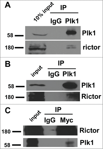
Figure 2. Plk1 phosphorylates Rictor-S1162 in vitro. (A) Recombinant Plk1 was incubated with 6 purified GST-Rictor regions [amino acidsCitation32 1–300, 301–600, 601–900, 901–1200, 1201–1500, 1501–1709] in the presence of [γ-32P]ATP. The reaction mixtures were resolved by SDS-PAGE, stained with Coomassie brilliant blue (Coom.), and detected by autoradiography. (B) Plk1 was incubated with the indicated forms of GST-Rictor fragment (aa 901–1200, C1). (C) Alignment of Rictor protein sequences containing Ser1162 in different species.
![Figure 2. Plk1 phosphorylates Rictor-S1162 in vitro. (A) Recombinant Plk1 was incubated with 6 purified GST-Rictor regions [amino acidsCitation32 1–300, 301–600, 601–900, 901–1200, 1201–1500, 1501–1709] in the presence of [γ-32P]ATP. The reaction mixtures were resolved by SDS-PAGE, stained with Coomassie brilliant blue (Coom.), and detected by autoradiography. (B) Plk1 was incubated with the indicated forms of GST-Rictor fragment (aa 901–1200, C1). (C) Alignment of Rictor protein sequences containing Ser1162 in different species.](/cms/asset/a276aabf-55a6-46a8-a5af-00f1fbcf1bf8/kccy_a_998050_f0002_b.gif)
Figure 3. Plk1 phosphorylates Rictor-S1162 in variety of human cancer cell lines. (A) Plk1 was incubated with GST-Rictor-aa 901–1200 (WT or S1162A) in the presence of unlabelled ATP, followed by anti-pS1162-Rictor IB. (B and C) PC3 (B) and Panc1 (C) cells were treated with mimosine (Mimo), hydroxyurea,Citation26 or nocodazole (Noc) to arrest cells at G1, S or M phase, respectively, and harvested for IB. (D) DU145 cells were treated with nocodazole, BI2536 or combination of these 2 drugs and harvested for anti-pS1162-Rictor IP, followed by anti-Rictor IB. (E) HeLa cells were depleted of Plk1 with dsRNA, treated with nocodazole and immunoblotted. (F) Cos7 cells were transfected with Myc-Rictor and harvested for IB.
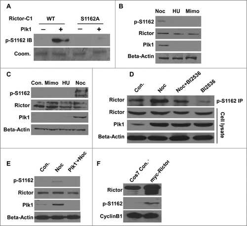
Figure 4. Plk1 phosphorylation of Rictor at S1162 does not regulate the mTOR pathway. (A) HEK 293T cells were transfected with various Myc-Rictor constructs (WT, S1162A or S1162E) and subjected to IB with antibodies against pS473-AKT and pT389-S6K1. (B) HEK 293T cells expressing various forms of Myc-Rictor were harvested for IB with indicated antibodies. (C) HEK 293T cells were transfected with Myc-Rictor (WT or S1162A), treated with LY294002 or rapamysin, and harvested for IB. A, S1162A; E, S1162E. (D) Cos7 cells were transfected with Myc-Rictor constructs and harvested for IB.
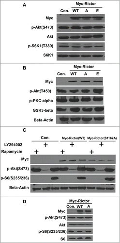
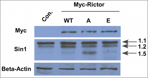
Figure 5. Phosphorylation of Rictor by Plk1 regulates the expression level of mSin1.5, but not other isoforms of mSin. HEK-293T cells were transfected with Myc-Rictor constructs (WT, S1162A or S1162E), and harvested for anti-mSin1 IB.