Figures & data
Figure 1. Cytoplasmic PTEN is functionally inhibited by BCR-ABL. (A) Representative Phosho-AKT immunofluorescence staining in primary CML Lin-CD34+CD38+ and Lin+ cells. PI: propidium iodide; P-AKT: phospho-Ser473-AKT. (B) Western immunoblot on Phospho-Ser473-AKT and Phospho-ERK in BCR-ABL-NIH3T3 cells. (C) Representative phosphatidylinositol (3,4,5)-triphosphate (PIP3) staining immunofluorescence in CD34 normal bone marrow and CML sample; PIP3 immunofluorescence staining intensity quantification with ImageJ software in 25 cells per conditions. (D) Upper panel: western immunoblot of primary total bone marrow CML extracts. Phospho-PTEN: Phospho-S380/T382/T383. Lower panel: quantification of Phospho-PTEN/PTEN in CML cases #1,#2,#3 of the western presented in the upper panel and CML cases #4,#5,#6 shown in the Supplementary B compared to the 2 normal bone marrow cases; *P <0.05. (E) Evaluation of PTEN activity in CML cases #1,#2,#3 of panel (d) and 2 normal bone marrow samples. A.U.: arbitrary unity; *P<0.05.
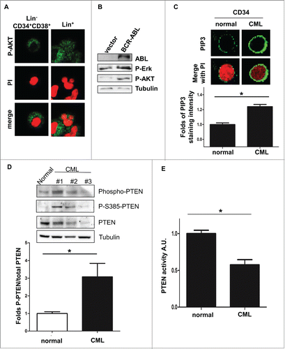
Figure 2. BCR-ABL promotes PTEN inactivation through Casein Kinase II. (A) PTEN-phosphorylation in parental and BCR-ABL-NIH3T3 treated with 1 μM imatinib and 60 μM TBB for 10 hours. To obtain a comparable level of expression of PTEN, the amount of BCR-ABL-NIH3T3 extracts were increased of 20% compared to parental NIH3T3 cells. (B) Schematic representation of PTEN with the indication of PTEN tail phosphorylation sites. (C) Co-immunoprecipitation of CKII-α and p210-BCR-ABL in BCR-ABL-infected NIH3T3 cells. (D) In vitro kinase assay with immunoprecipitated PTEN and CKII-α and purified ABL kinase. One μM Imatinib was added to the in vitro reaction for 10 minutes. Phospho-PTEN: Phospho-S380/T382/T383; P-Tyr: Phospho-Tyrosine: the band corresponds to the BCR-ABL molecular weight. (E) Transient transfection of Myc-PTEN mutants in BCR-ABL-infected NIH3T3 cells. 48 hours after transfection, myc-tag immunofluorescence was performed to verify PTEN compartmentalization. PTEN-TM: PTEN-S380A/T382A/T383A.
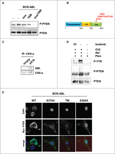
Figure 3. PTEN reactivation promotes apoptosis in BCR-ABL positive cells. (A) Parental and BCR-ABL-NIH3T3 were transfected with the indicated GFP-tagged-PTEN vectors. After 48 hours, PTEN phosphatase assay was performed in immunoprecipitated GFP-PTEN. *P <0.05; **P<0.01. (B) Western immunoblot of BCR-ABL-NIH3T3 cells transfected with the indicated GFP-PTEN mutants. Phospho-PTEN: PTEN-S380/T382/T383. (C) Apoptosis was assessed in NIH3T3 cells treated with 60 μM TBB and 1 μM Imatinib for 10 hours. Data are mean and d.s. of two independent experiments. After short incubation period with inhibitors, in BCR-ABL-NIH3T3 cells, solvent vs. TBB and solvent vs. TBB/imatinib differences are significant with *P<0.05.
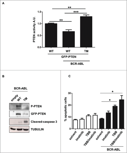
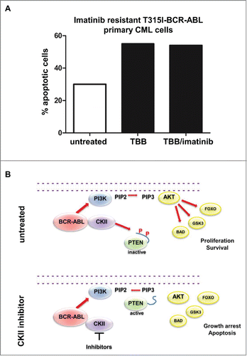
Figure 4. CKII inhibitor promotes apoptosis in T315I-BCR-ABL positive cells. (A) Apoptosis induction of BCR-ABL-T315I bone marrow sample treated with 60 μM TBB and 1 μM Imatinib for 10 hours. (B) Model of the BCR-ABL/CKII/PTEN network in CML.