Figures & data
Figure 1. Conditional senescence in SVts8 cells. (A) Representative images of SVts8 cells at the permissive (34°C) and non-permissive (39°C) temperatures, stained with DAPI or for SA-βgal activity, CCND1, CDK4 and γH2AX as indicated. (B) Cell size measurements of SVts8 cells grown at 34°C or switched to 39°C for 5 days. (C) Cell cycle profiles of SVts8 cells grown at 34°C or switched to 39°C for 5 days. The percentage of cells in the G1, S, and G2/M phases and with a >4N DNA content are indicated in each panel.
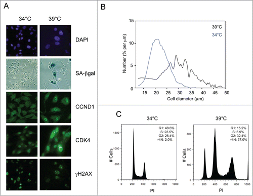
Figure 2. Status of cell cycle regulatory components at the permissive and non-permissive temperatures. (A) SVts8 cells cultured in the presence and absence of PD0332991 were switched from 34°C to 39°C for 6 d and cell lysates were fractionated by SDS-PAGE and immunoblotted with antibodies that detect specific phosphorylation events on pRb. MEK was used as a loading control. (B) Similar analysis of BJ-TERT-tsLT cells 6 d after temperature shift with antibodies against cyclin A (CCNA), p53, p21CIP1 and CDK4. (C) Effects of temperature shift on p21CIP1 and p16INK4a levels in SVts8 cells. (D) SVts8 cells at 34°C were infected with a retrovirus encoding shRNA against p21CIP1 (+) or an empty vector control (−). After antibiotic selection, cells were replated and grown at 34°C or 39°C for 48 h. Equivalent amounts of cell lysate were either analyzed directly by immunoblotting for p21CIP1, p16INK4a and MEK or immunoprecipitated with an antibody against CDK2 and assayed for their ability to phosphorylate GST-pRb in the presence of [γ-32P]ATP. (E) Phosphorylation status of pRb in SVts8 cells transduced with p21CIP1 or control shRNA. Equivalent amounts of cell lysate were analyzed directly by immunoblotting with the indicated antibodies. (F) SVts8 cells transduced with p21CIP1-specific shRNA continue to grow at 39°C for at least 7 d The plots record cell numbers relative to those before the temperature shift.
![Figure 2. Status of cell cycle regulatory components at the permissive and non-permissive temperatures. (A) SVts8 cells cultured in the presence and absence of PD0332991 were switched from 34°C to 39°C for 6 d and cell lysates were fractionated by SDS-PAGE and immunoblotted with antibodies that detect specific phosphorylation events on pRb. MEK was used as a loading control. (B) Similar analysis of BJ-TERT-tsLT cells 6 d after temperature shift with antibodies against cyclin A (CCNA), p53, p21CIP1 and CDK4. (C) Effects of temperature shift on p21CIP1 and p16INK4a levels in SVts8 cells. (D) SVts8 cells at 34°C were infected with a retrovirus encoding shRNA against p21CIP1 (+) or an empty vector control (−). After antibiotic selection, cells were replated and grown at 34°C or 39°C for 48 h. Equivalent amounts of cell lysate were either analyzed directly by immunoblotting for p21CIP1, p16INK4a and MEK or immunoprecipitated with an antibody against CDK2 and assayed for their ability to phosphorylate GST-pRb in the presence of [γ-32P]ATP. (E) Phosphorylation status of pRb in SVts8 cells transduced with p21CIP1 or control shRNA. Equivalent amounts of cell lysate were analyzed directly by immunoblotting with the indicated antibodies. (F) SVts8 cells transduced with p21CIP1-specific shRNA continue to grow at 39°C for at least 7 d The plots record cell numbers relative to those before the temperature shift.](/cms/asset/69f91b0c-1017-4288-8883-b3536dce6e12/kccy_a_1010866_f0002_b.gif)
Figure 3. Comparison of cyclin-CDK-CKI interactions at the permissive and non-permissive temperatures. (A) Cell lysates were prepared from SVts8 (SV) or BJ-TERT-tsLT (BJ) cells grown at 34°C or 6 d after shifting to 39°C. Equal amounts of protein were either analyzed directly by SDS-PAGE and immunoblotting (left panel) or after immunoprecipitation with an antibody against CDK4 (right panel). (B) Equal amounts of cell lysate (300 μg of total protein) from SVts8 cells grown at 34°C and 39°C were immunoprecipitated with antisera against CCND1, CDK4, p21CIP1 and p16INK4a as indicated. After fractionation by SDS-PAGE, the precipitated proteins were immunoblotted for CCND1, CDK4, p21CIP1 and p16INK4a as indicated. The total amounts of CCND1, CDK4, p21CIP1 and p16INK4a were monitored by direct immunoblotting of 30 μg of each protein sample (Total). (C) Similar analysis of CCND1 immunoprecipitates from SVts8 cells transduced with an shRNA against p21CIP1 or non-specific shRNA.
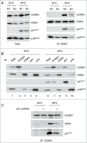
Figure 4. Effects of CDK4 knockdown on the senescence phenotype. (A) Representative images of SVts8 cells transduced with CDK4 or control shRNA at the permissive (34°C) and non-permissive (39°C) temperatures. Cells were stained with DAPI or for CDK4 as indicated. (B) Cell size measurements of SVts8 cells transduced with CDK4 or control shRNA at 34°C or switched to 39°C for 6 days. (C) Cell cycle profiles of SVts8 cells transduced with CDK4 shRNA or control shRNA at 34°C, or switched to 39°C for 5 days.
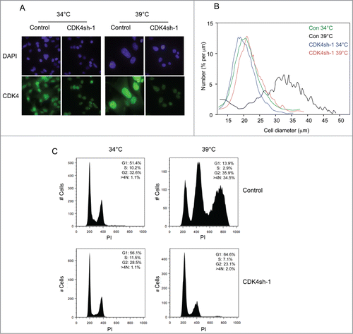
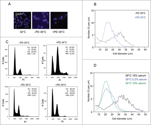
Figure 5. Effects of CDK4 inhibition on the senescence phenotype. (A) Representative images of SVts8 cells at the permissive temperature (34°C) or after shifting to the non-permissive temperature (39°C) in the presence or absence of the CDK4 inhibitor PD0332991 (4μM). The cells were stained with DAPI. (B) Mean cell size comparisons of SVts8 at the non-permissive temperature in the presence or absence of the CDK4 inhibitor PD0332991. (C) Cell cycle profiles of SVts8 cells at 34°C or 39°C in the presence or absence of the CDK4 inhibitor PD0332991. (D) Mean cell size comparisons of SVts8 at 34°C or after shifting to 39°C in either 0.2% or 10% serum.