Figures & data
Figure 1. (A) Schematic of the separated mouse vibrissa hair follicle. The upper part of the vibrissa hair follicle contains nestin-expressing, CD34-positive, K15-negative hair-follicle-associated-pluripotent (HAP) stem cells. (B) The mouse vibrissa hair follicle was separated into 3 parts (upper, middle, and lower part). Each hair follicle part was separately suspended in DMEM containing 10% FBS. (C) Two weeks after culture all 3 parts (upper, middle, and lower) of the hair follicle, incubated in DMEM with 10% FBS cells differentiated to troponin- and desmin-positive cardiac muscle cells. The number of cardiac muscle cells was significantly higher in the upper part compared to the middle, and lower part of the hair follicle. Bars 100 μm.
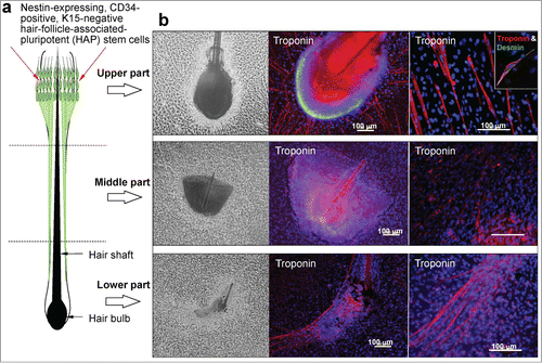
Figure 2. (A) Total cell number differentiated from the upper, middle and lower part of the hair follicle. (B) The number of cardiac muscle cells differentiated from the upper, middle and lower parts of the hair follicle. *P < 0.01 vs upper part.
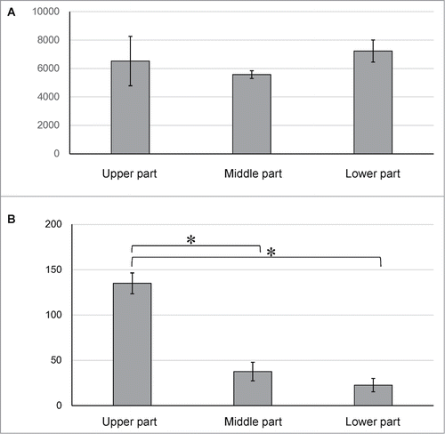
Figure 3. Fluorescence-activated cell sorting (FACS) analysis showed that all 3 parts of the hair follicle differentiated to troponin-positive cardiac muscle cells, βIII-tubulin-positive neurons, K15-positive keratinocytes, smooth muscle actin-positive cells, and GFAP-positive glial cells.
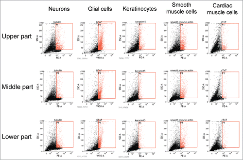
Table 1. Percentage of cardiac muscle cells and other cell types differentiate from the separated upper, middle and lower parts of the mouse whisker follicle
Figure 4. Effect of isoproterenol and propranolol on the beat rate of cardiac muscle cells differentiated from the upper part of the hair follicle. * P < 0.01 vs control, †P < 0.01 vs isoproterenol.
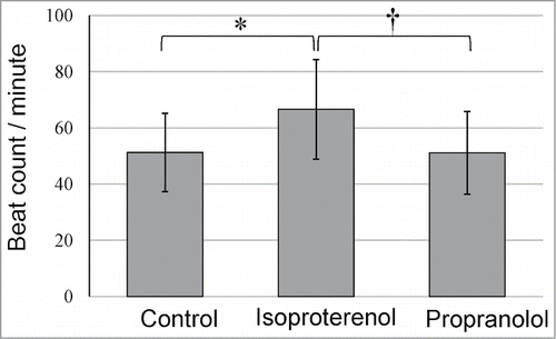
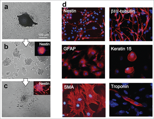
Figure 5. (A) The upper part of hair follicle was cultured for 4 weeks in DMEM with 10% FBS. (B) Cells growing out from the upper part of the hair follicle were transferred to DMEM/F12 without FBS. Two weeks later, the growing cells formed many nestin-expressing hair spheres. (C) Two days after transfer to DMEM with 10% FBS, the hair spheres started to differentiate. (D) One week after switching to DMEM containing 10% FBS, the hair spheres differentiated to desmin-positive cardiac muscle cells, nestin- and βIII-tubulin-positive neurons, GFAP-positive glial cells, K15-positive keratinocytes, and actin-positive cells.