Figures & data
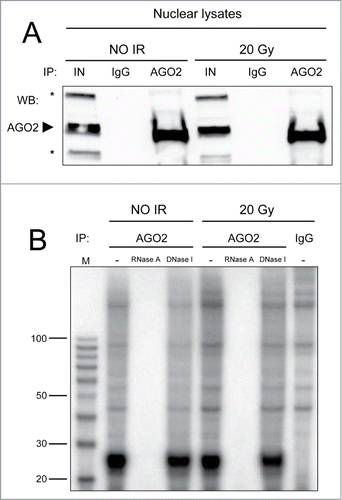
Figure 1. (A) AGO2 IP efficiency was tested by Western blot. Five% of each AGO2 IP (lanes AGO2) or mock IP (lanes IgG), performed in nuclear extracts of irradiated (20 Gy) or not irradiated (NO IR) HeLa cells, was resolved onto a 4–15% SDS-PAGE along with 1% of the input lysates (lanes IN). Proteins were transferred onto nitrocellulose membrane and probed for AGO2. The arrowhead indicates endogenous AGO2, whereas the asterisks mark unspecific bands. No AGO2 is retained in the mock IPs. (B) Human AGO2 binds to small RNAs only and not to small DNAs in the nucleus. AGO2 co-precipitated nucleic acids (lanes AGO2) from irradiated (20 Gy) or not irradiated (NO IR) HeLa nuclei were 5′-radioactively labeled, treated with RNase A or DNase I and fractionated onto a 10% urea PAGE along with a size marker (lane M). As controls, both untreated samples (lanes -) and nucleic acids from mock IP (lane IgG) were also fractionated.