674
Views
1
CrossRef citations to date
0
Altmetric
NEWS & VIEWS

Atrial myocytes demonstrate the diversity of cardiac calcium signalling

&
Pages 219-220 | Received 04 Aug 2015, Accepted 20 Aug 2015, Published online: 05 Nov 2015

Figures & data

Figure 1. The Figure shows an illustration of part of an atrial myocyte. The arrows denote the movement of Ca2+ ions as a centripetal wave. The Ca2+ wave propagates via CICR between neighbouring clusters of RyRs. In the presence of InsP3, Ca2+ wave propagation is boosted by the opening of InsP3Rs. Both Ca2+ and InsP3 can pass through nuclear pore complexes (NPC), and trigger nucleoplasmic Ca2+ signals that influence gene transcription. Mitochondria sequester Ca2+ ions, and thereby retard Ca2+ wave propagation.
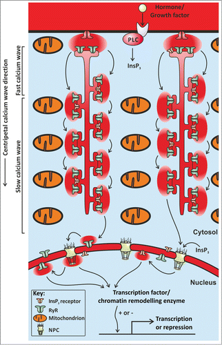
Figure 1. The Figure shows an illustration of part of an atrial myocyte. The arrows denote the movement of Ca2+ ions as a centripetal wave. The Ca2+ wave propagates via CICR between neighbouring clusters of RyRs. In the presence of InsP3, Ca2+ wave propagation is boosted by the opening of InsP3Rs. Both Ca2+ and InsP3 can pass through nuclear pore complexes (NPC), and trigger nucleoplasmic Ca2+ signals that influence gene transcription. Mitochondria sequester Ca2+ ions, and thereby retard Ca2+ wave propagation.