927
Views
4
CrossRef citations to date
0
Altmetric
Report

CHFR is important for the survival of male premeiotic germ cells

&
Pages 3454-3460 | Received 29 Apr 2015, Accepted 07 Sep 2015, Published online: 13 Nov 2015

Figures & data

Figure 1. A large portion of Chfr knockout male mice are infertile. (a) Wild type (WT) and Chfr knockout (KO) male mice were mated with WT female mice. Male mice of 3 different ages were used. The numbers of fertile and infertile mice are shown. The age at the start and the end of mating are marked. (b) Sperm were harvested from epididymides at the end of mating. Sperm counts are shown. (c) Hematoxylin and eosin staining of epididymis sections are shown. Higher magnifications of selected areas are shown on right. Scale bar, 50 µM.
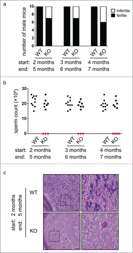
Figure 1. A large portion of Chfr knockout male mice are infertile. (a) Wild type (WT) and Chfr knockout (KO) male mice were mated with WT female mice. Male mice of 3 different ages were used. The numbers of fertile and infertile mice are shown. The age at the start and the end of mating are marked. (b) Sperm were harvested from epididymides at the end of mating. Sperm counts are shown. (c) Hematoxylin and eosin staining of epididymis sections are shown. Higher magnifications of selected areas are shown on right. Scale bar, 50 µM.

Figure 2. Germ cells are completely lost in small testes from Chfr knockout male mice at 12 weeks. (a) Typical pictures of normal testes from WT male mice and small testes from Chfr knockout male mice are shown. (b) The weight of normal testes from WT male mice and small testes from Chfr knockout male mice are shown. (c) Sperm were harvested from epididymides of mice shown in (b), and sperm counts are shown. (d) Testis sections of 12-week-old WT and Chfr knockout male mice were stained with periodic acid schiff (PAS)-hematoxylin. Typical pictures are shown. Scale bar, 50 µM. Asterisk: large vacuoles. (e-f) Seminiferous tubules with germ cells (e) and with large vacuoles (f) are summarized. Mean and standard deviation are shown.
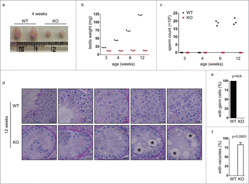
Figure 2. Germ cells are completely lost in small testes from Chfr knockout male mice at 12 weeks. (a) Typical pictures of normal testes from WT male mice and small testes from Chfr knockout male mice are shown. (b) The weight of normal testes from WT male mice and small testes from Chfr knockout male mice are shown. (c) Sperm were harvested from epididymides of mice shown in (b), and sperm counts are shown. (d) Testis sections of 12-week-old WT and Chfr knockout male mice were stained with periodic acid schiff (PAS)-hematoxylin. Typical pictures are shown. Scale bar, 50 µM. Asterisk: large vacuoles. (e-f) Seminiferous tubules with germ cells (e) and with large vacuoles (f) are summarized. Mean and standard deviation are shown.

Figure 3. Germ cells are decreased in small testes from Chfr knockout male mice at 6 weeks. Testis sections of 12-week-old male mice were stained with periodic acid schiff (PAS)-hematoxylin. Typical pictures of normal testes from WT male mice (a-e) and small testes from Chfr knockout male mice (f-o) are shown. Scale bar, 50 µM. Asterisk: large vacuoles. (p-r) Seminiferous tubules with germ cells (p), with multiple layers of germs cells (q), and with large vacuoles (r) are summarized. Mean and standard deviation are shown.
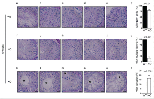
Figure 3. Germ cells are decreased in small testes from Chfr knockout male mice at 6 weeks. Testis sections of 12-week-old male mice were stained with periodic acid schiff (PAS)-hematoxylin. Typical pictures of normal testes from WT male mice (a-e) and small testes from Chfr knockout male mice (f-o) are shown. Scale bar, 50 µM. Asterisk: large vacuoles. (p-r) Seminiferous tubules with germ cells (p), with multiple layers of germs cells (q), and with large vacuoles (r) are summarized. Mean and standard deviation are shown.

Figure 4. Spermatogenesis in Chfr knockout male mice with small testes are delayed at 3 and 4 weeks. Testis sections of 3 and 4-week-old male mice were stained with periodic acid schiff (PAS)-hematoxylin. Typical pictures of normal testes from WT male mice (a-d for 4 weeks and k-m for 3 weeks) and small testes from Chfr knockout male mice (e-h for 4 weeks and n-p for 3 weeks) are shown. Scale bar, 50 µM. Asterisk: large vacuoles. (i-j) For 4-week-old male mice, seminiferous tubules with multiple layers of germs cells (i) and with large vacuoles (j) are summarized. Mean and standard deviation are shown.
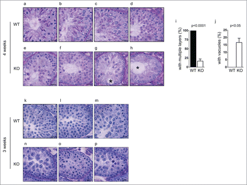
Figure 4. Spermatogenesis in Chfr knockout male mice with small testes are delayed at 3 and 4 weeks. Testis sections of 3 and 4-week-old male mice were stained with periodic acid schiff (PAS)-hematoxylin. Typical pictures of normal testes from WT male mice (a-d for 4 weeks and k-m for 3 weeks) and small testes from Chfr knockout male mice (e-h for 4 weeks and n-p for 3 weeks) are shown. Scale bar, 50 µM. Asterisk: large vacuoles. (i-j) For 4-week-old male mice, seminiferous tubules with multiple layers of germs cells (i) and with large vacuoles (j) are summarized. Mean and standard deviation are shown.

Figure 5. Apoptosis occur in germ cells from Chfr knockout male mice. (a-b) Apoptosis was detected in testis sections of 3 and 4-week-old male mice. Apoptotic cells are marked by arrow heads. Seminiferous tubules with apoptotic cells are summarized. Scale bar, 50 µM. (c) Levels of ATM, phosphorylated ATM at serine 1981, Chk2, and phosphorylated Chk2 are shown in WT and Chfr knockout MEFs with or without ionizing radiation (IR). (d) Levels of ATM, phosphorylated ATM at serine 1981, Chk2, and phosphorylated Chk2 are shown in 3-week-old WT and Chfr knockout testes. N, normal size testes. S, small testes. Quantification was performed. Mean and standard deviation are shown.
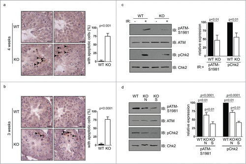
Figure 5. Apoptosis occur in germ cells from Chfr knockout male mice. (a-b) Apoptosis was detected in testis sections of 3 and 4-week-old male mice. Apoptotic cells are marked by arrow heads. Seminiferous tubules with apoptotic cells are summarized. Scale bar, 50 µM. (c) Levels of ATM, phosphorylated ATM at serine 1981, Chk2, and phosphorylated Chk2 are shown in WT and Chfr knockout MEFs with or without ionizing radiation (IR). (d) Levels of ATM, phosphorylated ATM at serine 1981, Chk2, and phosphorylated Chk2 are shown in 3-week-old WT and Chfr knockout testes. N, normal size testes. S, small testes. Quantification was performed. Mean and standard deviation are shown.

Reprints and Corporate Permissions

Please note: Selecting permissions does not provide access to the full text of the article, please see our help page How do I view content?

To request a reprint or corporate permissions for this article, please click on the relevant link below:

Academic Permissions

Please note: Selecting permissions does not provide access to the full text of the article, please see our help page How do I view content?

Obtain permissions instantly via Rightslink by clicking on the button below:

If you are unable to obtain permissions via Rightslink, please complete and submit this Permissions form. For more information, please visit our Permissions help page.