Figures & data
Figure 1 (See previous page). IL-12 but not IL-21 induced the differentiation of human naive CD4+ (T)cells into Th1 and Tfh co-expressing cells. Human naive CD4+ T cells from cord blood were stimulated for 5 d with or without anti-CD3 and anti-CD28 mAbs in the presence or absence of IL-12, IL-21 or IL-12 plus IL-21. The cells were harvested and rested for 2 d with IL-2. The cells were re-stimulated for 6 h with PMA and ionomycin in the presence of BFA. The expression of IL-21 and IFN-γ was detected by FACS. A representative result of different cytokines on the development of Th1 and Tfh cells was shown (A). Statistical data of percentage of IL-21+CD4+ (B), IFN-γ+CD4+ (C), IFN-γ−IL-21+ (D), IFN-γ−IL-21+ (E) and IFN-γ+IL-21+ (F) CD4+ T cells were mean ± SD from 5 independent experiments as described in A. Cell surface expression of CXCR5, PD-1, ICOS and CXCR3 was assessed by surface staining and a representative result was shown (G). P < 0 .05 was considered significant.
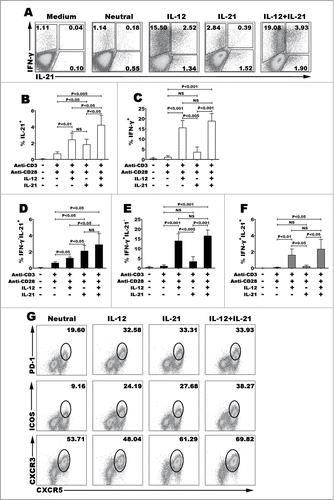
Figure 2. IL-12 induced the differentiation of polyfunctional CD4+ (T)cells. Naive CD4+ T cells were stimulated for 5 d with anti-CD3 and anti-CD28 mAbs in the presence of IL-12. The cells were rested and re-stimulated for 6 h with PMA and ionomycin in the presence of BFA. The expression of IL-21, IFN-γ, TNF-α and IL-2 was detected by FACS. Most of IL-21-expressing CD4+ T cells co-expressed Th1 cytokine IFN-γ, TNF-α or IL-2. The representative dot plots were shown (A). Gated on IL-21− and IL-21+CD4+ T cells or IFN-γ− and IFN-γ+CD4+ T cells, the expression of IFN-γ or IL-21 was analyzed. The representative histogram graphs and statistical data of mean MFI were shown (B). Gated on IL-21−IFN-γ−, IL-21+IFN-γ+, IL-21+IFN-γ− and IL-21−IFN-γ+ CD4+ T cells, the expression of TNF-α and IL-2 in the 4 different subsets were analyzed. The representative dot plots (C) and statistical data of mean percentages (D) of TNF-α−IL-2−, TNF-α−IL-2+, TNF-α+IL-2− and TNF-α+IL-2+ were shown.
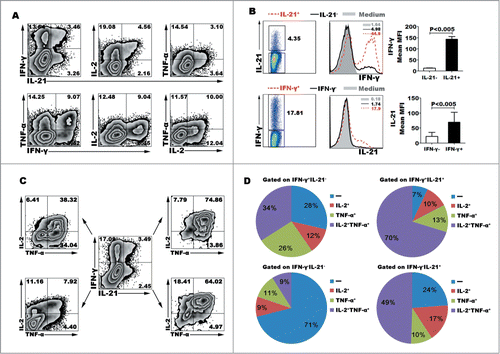
Figure 3. Kinetic studies of the expression of cytokines and transcription factors after IL-12 stimulation. Naive CD4+ T cells were stimulated for 1 to 5 d with anti-CD3 and anti-CD28 mAbs in the presence or absence of IL-12, and the cells and supernatants were harvested at different time-points. The levels of IFN-γ and IL-21 in supernatants were determined by ELISA (A). The cells were re-stimulated for 48 h with PMA and ionomycin, and levels of IFN-γ and IL-21 in supernatants were determined by ELISA (B). The cells were re-stimulated for 6 h with PMA and ionomycin in the presence of BFA, and the expression of IFN-γ, IL-21, T-bet and BCL−6 was detected by FACS. The representative results were shown (C, D).
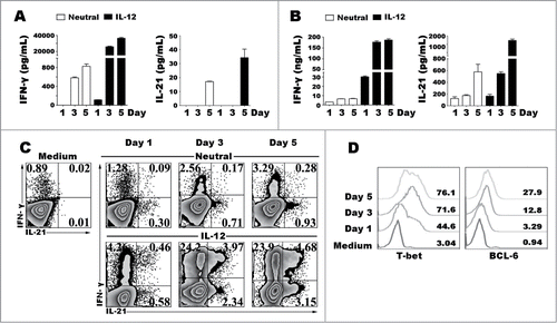
Figure 4. Characteristic phenotypes of Th1 and Tfh cells. Naive CD4+ T cells were stimulated for 5 d with anti-CD3 and anti-CD28 mAbs in the presence of IL-12. The cells were harvested, rested and re-stimulated for 6 h with PMA and ionomycin in the presence of BFA. The expression of cytokines and surface markers were detected by FACS. A representative result of surface expression of CXCR5, CXCR3, PD-1, ICOS and CD40L (A), and expression of transcription factor T-bet and BCL−6 (B) on IFN-γ−IL-21−, IFN-γ+IL-21−, IFN-γ−IL-21+ and IFN-γ+IL-21+ CD4+ T cells were shown in histogram, statistical data of MFI (B) were mean±SD from 5 independent experiments. A representative result of IL-21 and IFN-γ expression gated on T-bet−BCL−6−, T-bet−BCL−6+, T-bet+BCL−6+ and T-bet+BCL−6−CD4+ T cells was shown in histogram graph and dot plots (C). The statistical data were mean ± SD from 5 independent experiments (D).
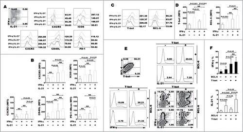
Figure 5. Effect of IFN-γ on modulating the expression and production of IL-21 and IFN-γ. Naive CD4+ T cells were stimulated for 3 d with anti-CD3 and anti-CD28 mAbs plus IL-12, and anti-IFN-γ or IFN-γ was added into the culture in the presence of IL-12 at day 0 and day 3, the expression of IL-21 and IFN-γ production were detected by FACS after re-stimulated with PMA and ionomycin at day 5, a representative dot plots were shown (A). Transcription factors phosphorylated STAT-1 and STAT-4, T-bet and BCL−6 were detected by FACS at day 3, a representative histogram graphs were shown (B). Naive CD4+ T cells were stimulated for 3 d in the presence of IL-12, IL-21 or IL-12 plus IL-21 with or without anti-IFN-γ. The cells were harvested, rested and re-stimulated for 6 h with PMA and ionomycin in the presence of BFA. IFN-γ and IL-21 production were analyzed by FACS (C). The cells were re-stimulated for 48 h with PMA and ionomycin, the levels of IFN-γ and IL-21 in supernatants were determined by ELISA (D). The cells were re-stimulated for 48 h with PMA and ionomycin in the presence of anti-IFN-γ. The levels of IFN-γ and IL-21 in supernatants were determined by ELISA (E). The expression of phenotypic markers (F) and transcription factor T-bet and BCL−6 (G) were analyzed by FACS under the conditions of IL-12 or IL-12 plus anti- IFN-γ. The representative histogram graphs and statistical data from 5 independent experiments were shown.
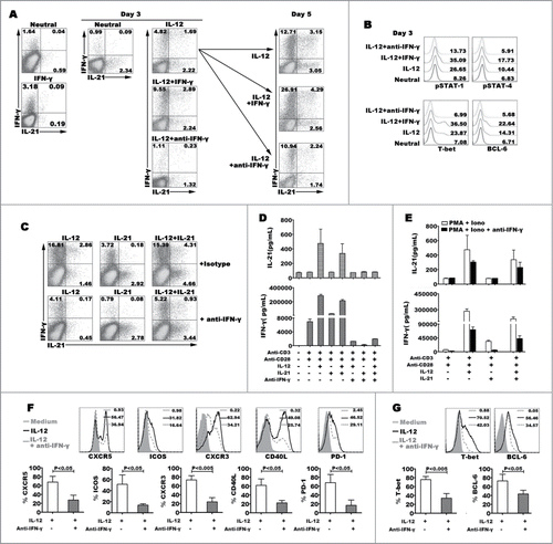
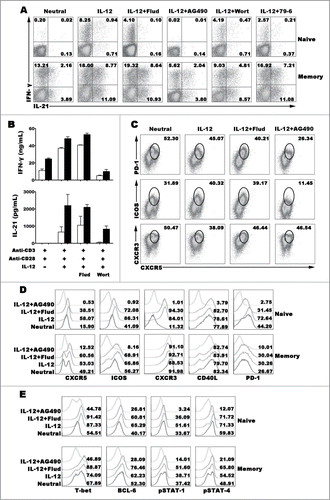
Figure 6 (See previous page). Transcription factors regulating the expression and production of IFN-γ and IL-21. Naive CD4+ T cells (CD4+CD45RA+CD45RO−) and memory CD4+ T cells (CD4+CD45RA−CD45RO+) were stimulated with anti-CD3 and anti-CD28 plus IL-12 in the presence or absence of inhibitors for transcription factor for 5 d. The cells were harvested and re-stimulated with PMA and ionomycin for 6 h in the presence of BFA, and the expression of IFN-γ and IL-21 were detected by FACS (A). Cells were re-stimulated with PMA and ionomycin for 48 h, the levels of IFN-γ and IL-21 in the supernatants were detected by ELISA (B). Memory CD4+ T cells were stimulated with anti-CD3 and anti-CD28 plus IL-12 in the presence or absence of inhibitors for transcription factor for 5 d. The cells were harvested and re-stimulated with PMA and ionomycin for 6 h in the presence of BFA, and the expression of IFN-γ, IL-21, CXCR5, CXCR3, ICOS and PD-1 were detected by FACS (C). Expression of Tfh and Th1 cell phenotypes (D) and transcription factors (E) was detected by FACS.