2,067
Views
27
CrossRef citations to date
0
Altmetric
Report

The oncolytic peptide LTX-315 triggers necrotic cell death

, , , , , , , , , & show all
Pages 3506-3512 | Received 31 Jul 2015, Accepted 07 Sep 2015, Published online: 13 Nov 2015

Figures & data

Figure 1. Ultrastructural characteristics of LTX-315-induced cell death. U2OS cells were either left untreated (control, Ctr) or treated with the indicated dose of LTX-315 for 6 hours followed by osmium tetroxide staining and transmission electron microscopy. Note the presence of dilated mitochondria in cells treated with 12.5 or 50 µg/ml of LTX-315.
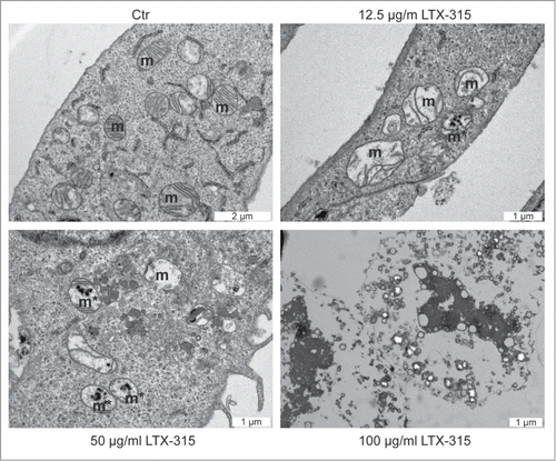
Figure 1. Ultrastructural characteristics of LTX-315-induced cell death. U2OS cells were either left untreated (control, Ctr) or treated with the indicated dose of LTX-315 for 6 hours followed by osmium tetroxide staining and transmission electron microscopy. Note the presence of dilated mitochondria in cells treated with 12.5 or 50 µg/ml of LTX-315.

Figure 2. Failure of LTX-315 to induce caspase-3 activation and nuclear shrinkage. U2OS cells were treated for the indicated period with LTX-315, staurosporine (STS) or 100 µM carbonyl cyanide m-chlorophenyl hydrazine (CCCP) in the absence or presence of the pan-caspase inhibitor Z-VAD or the necroptosis inhibitor necrostatin-1 (Nec), followed by fixation and permeabilization of the cells, immunofluorescence staining for the detection of active caspase 3 (Casp3a) and counterstained with the chromatin dye Hoechst 33342. Representative images are shown in A (images obtained in the absence of Z-VAD and Nec). Quantitative results (means ± SD of triplicates) are shown in B and C. In B, the frequency of Casp3a+ cells is shown for each treatment, while in C, the frequency of cells with normal morphology (not shrunken) is displayed. Asterisks indicate significant differences (unpaired Student t test) with respect to untreated controls. *p < 0.05; **p < 0.01; ***p < 0.001.
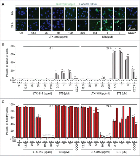
Figure 2. Failure of LTX-315 to induce caspase-3 activation and nuclear shrinkage. U2OS cells were treated for the indicated period with LTX-315, staurosporine (STS) or 100 µM carbonyl cyanide m-chlorophenyl hydrazine (CCCP) in the absence or presence of the pan-caspase inhibitor Z-VAD or the necroptosis inhibitor necrostatin-1 (Nec), followed by fixation and permeabilization of the cells, immunofluorescence staining for the detection of active caspase 3 (Casp3a) and counterstained with the chromatin dye Hoechst 33342. Representative images are shown in A (images obtained in the absence of Z-VAD and Nec). Quantitative results (means ± SD of triplicates) are shown in B and C. In B, the frequency of Casp3a+ cells is shown for each treatment, while in C, the frequency of cells with normal morphology (not shrunken) is displayed. Asterisks indicate significant differences (unpaired Student t test) with respect to untreated controls. *p < 0.05; **p < 0.01; ***p < 0.001.

Figure 3. LTX-315-induced cell death is refractory to inhibition by Z-VAD and necrostatin 1. U2OS cells were treated for the indicated period with LTX-315, staurosporine (STS) or 100 µM carbonyl cyanide m-chlorophenyl hydrazine (CCCP) in the absence or presence of the pan-caspase inhibitor Z-VAD or the necroptosis inhibitor necrostatin-1 (Nec), followed by staining with the vital dye propidium iodine (PI) and the chromatin dye Hoechst 33342. Representative fluorescence microphotographs (images obtained in the absence of Z-VAD and Nec) are shown in A, and quantitative results are show in B. Asterisks indicate significant differences (unpaired Student t test) in Hoechstdim PI- cell number with respect to untreated controls, for a given co-treatment. *p < 0.05; **p < 0.01; ***p < 0.001. Sharps indicate significant differences, for a given drug treatment, compared to control without co-treatment. #p < 0.05; ##p < 0.01.
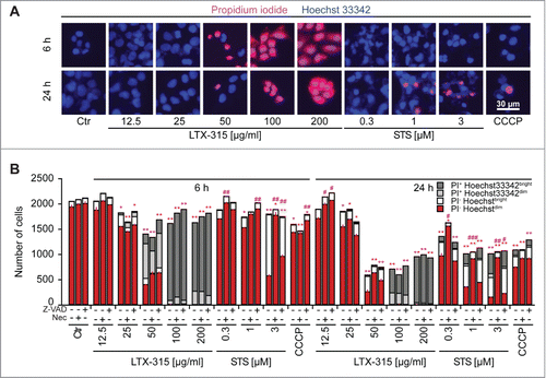
Figure 3. LTX-315-induced cell death is refractory to inhibition by Z-VAD and necrostatin 1. U2OS cells were treated for the indicated period with LTX-315, staurosporine (STS) or 100 µM carbonyl cyanide m-chlorophenyl hydrazine (CCCP) in the absence or presence of the pan-caspase inhibitor Z-VAD or the necroptosis inhibitor necrostatin-1 (Nec), followed by staining with the vital dye propidium iodine (PI) and the chromatin dye Hoechst 33342. Representative fluorescence microphotographs (images obtained in the absence of Z-VAD and Nec) are shown in A, and quantitative results are show in B. Asterisks indicate significant differences (unpaired Student t test) in Hoechstdim PI- cell number with respect to untreated controls, for a given co-treatment. *p < 0.05; **p < 0.01; ***p < 0.001. Sharps indicate significant differences, for a given drug treatment, compared to control without co-treatment. #p < 0.05; ##p < 0.01.

Figure 4. Failure of cyclosporine A to inhibit LTX-315-induced cell death. U2OS cells were treated for 6 hrs with LTX-315 in the absence or presence of 100 nM of the mitochondrial permeability transition inhibitor cyclosporine A (CsA), followed by staining with the vital dye propidium iodine (PI) and the chromatin dye Hoechst 33342. Representative fluorescence microphotographs are shown in A, and quantitative results are shown in B. Asterisks indicate significant differences (unpaired Student t test) in Hoechstdim PI- cell number with respect to untreated controls, for a given co-treatment. *p < 0.05; **p < 0.01; ***p < 0.001.
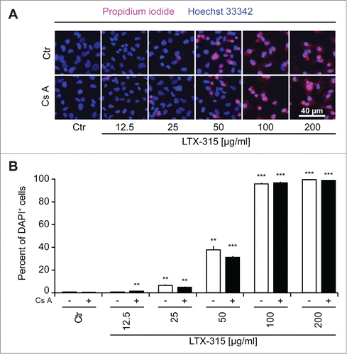
Figure 4. Failure of cyclosporine A to inhibit LTX-315-induced cell death. U2OS cells were treated for 6 hrs with LTX-315 in the absence or presence of 100 nM of the mitochondrial permeability transition inhibitor cyclosporine A (CsA), followed by staining with the vital dye propidium iodine (PI) and the chromatin dye Hoechst 33342. Representative fluorescence microphotographs are shown in A, and quantitative results are shown in B. Asterisks indicate significant differences (unpaired Student t test) in Hoechstdim PI- cell number with respect to untreated controls, for a given co-treatment. *p < 0.05; **p < 0.01; ***p < 0.001.

Figure 5. Videomicroscopy of cellular disintegration. U2OS cells were treated for the indicated period with LTX-315 in the presence of 1 µM DAPI. Morphological changes and the uptake of the exclusion dye were monitored by videomicroscopy. Representative image galleries from U2OS cells treated with 50 µg/ml LTX-315 or left untreated are depicted.

Figure 5. Videomicroscopy of cellular disintegration. U2OS cells were treated for the indicated period with LTX-315 in the presence of 1 µM DAPI. Morphological changes and the uptake of the exclusion dye were monitored by videomicroscopy. Representative image galleries from U2OS cells treated with 50 µg/ml LTX-315 or left untreated are depicted.

Reprints and Corporate Permissions

Please note: Selecting permissions does not provide access to the full text of the article, please see our help page How do I view content?

To request a reprint or corporate permissions for this article, please click on the relevant link below:

Academic Permissions

Please note: Selecting permissions does not provide access to the full text of the article, please see our help page How do I view content?

Obtain permissions instantly via Rightslink by clicking on the button below:

If you are unable to obtain permissions via Rightslink, please complete and submit this Permissions form. For more information, please visit our Permissions help page.