
Figures & data
Figure 1. Dose-response curves showing IC50 of hexane extract of Erythroxylum daphnites (EDH) on inhibition of both SCC-9 (tongue carcinoma cells) and HaCaT (human Keratinocyte cells) cell viability. Dose-response curves were assessed by MTT assay in SCC-9 (A) and HaCat cells (B) after 24 h of treatment with increased concentration of EDH. The results represent the percentage of dead cells in the presence of different doses of EDH. They are representative of at least 3 independent experiments in triplicate and show the mean ± SEM.
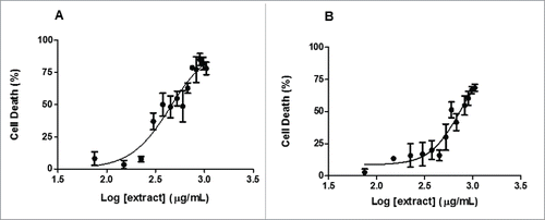
Figure 2. The hexane extract of Erythroxylum daphnites extract (EDH) decreases the proliferation of SCC-9 tongue carcinoma cells. Cell proliferation was measured using BrdU incorporation assay as described in methods. The treatment with EDH significantly decreased the proliferation of SCC-9 cells compared to control. *P < 0.01 vs. control (Student's t test).
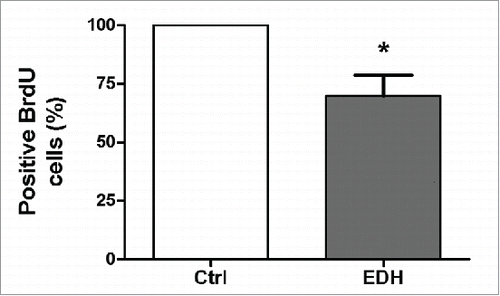
Figure 3. A – Distribution of cells in each stage of the cell cycle after 24 h of treatment with the hexane extract of Erythroxylum daphnites (EDH) (*P < 0.0001 vs. proportion of cells in S-phase). B - Distribution of events labeled with propidium iodide in the histogram, the first gray peak represents events in G0/G1 phase, the second gray peak (right) represents events in G2/M phase, and the hatched region represents the number of events in S phase.
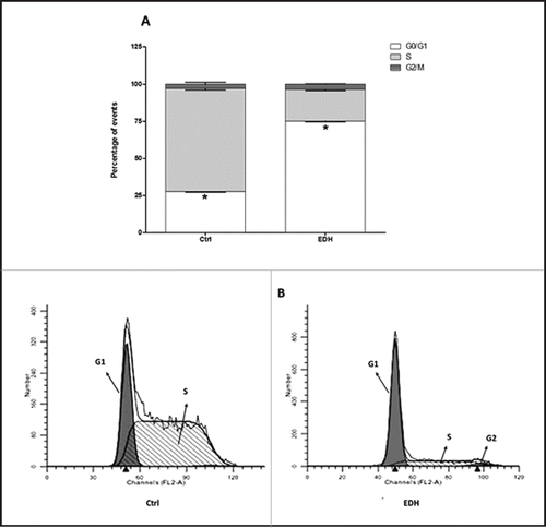
Figure 4. Expression of cell cycle proteins in SCC-9 tongue carcinoma cells assessed by Western blot after 24 hours of treatment with hexane extract of Erythroxylum daphnites (EDH). All these proteins were involved in the transition to G1/S phase of cell cycle. The reduction of expression of cyclin D, cyclin E and p27 and enhanced level of p21 suggest the arrest of cells treated with EDH at G0/G1 phase.
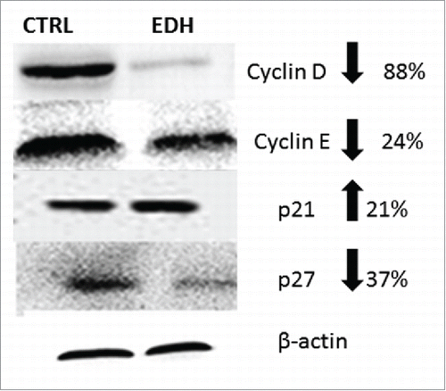
Figure 5. Expression of cleavage of Pro-caspase 3 and caspase in SCC-9 tongue carcinoma cells assessed by Western blot after 12 hours of treatment with hexane extract of Erythroxylum daphnites (EDH). The presence of caspase 3 band in SCC-9 cells EDH treated with EDH confirms an apoptotic effect. GAPDH expression was used as housekeeping control (Ctr). The figure represents one of 3 experiments.
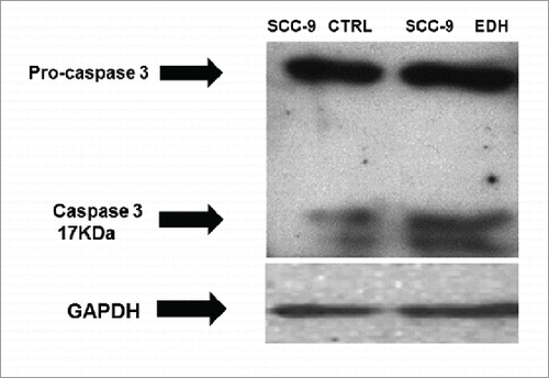
Figure 6. A - Cytotoxicity of hexane extract of Erythroxylum daphnites (EDH) fractions after 24 h of treatment of SCC-9 tongue carcinoma cells. 1 - Fraction hexane:ethyl acetate (1:1) (EDH-FHE); 2 - Fraction ethyl acetate:methanol (EDH-FEM) (1:1); 3 - Fraction ethyl acetate (EDH-FE); 4 - Fraction methanol (EDH-FM); 5 - Fraction hexane (EDH-FH). *P < 0.01 vs. control. B - Chemical structure of compounds found in fraction EDH-FHE. The results show the percentage of cell viability in the presence of different extracts treatment.
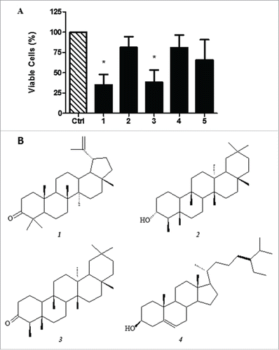
Table 1. Components identified in the fraction EDH-FHE by GC-MS analysis.
Table 2. List of primary antibodies.