Figures & data
Figure 1. Experimental scheme for subcutaneous transplantation of whisker hair follicles. Whisker hair follicles were first isolated from both young and old nestin-driven green fluorescent protein (ND-GFP) transgenic hairy mice and placed into culture medium. Both young and old hair follicles were subsequently transplanted into the subcutis on both flanks of young and old non-transgenic nude mice. Please see the Materials and Methods for details.
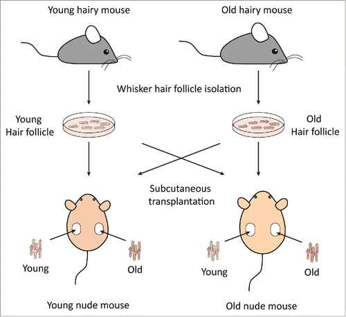
Figure 2. Comparsion of hair-shaft growth from young and old hair follicles transplanted in young or old nude mice. Whisker follicles were isolated from young and old ND-GFP transgenic hairy mice and transplanted to young and old non-transgenic nude mice. Hair-shaft length was measured with the Dino-Lite microscope imager. Please see Materials and Methods for details.
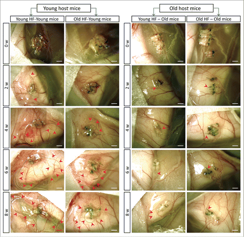
Figure 3. Quantitative time-course growth data of the average of the 10 longest hair shafts in the different groups after hair-follicle subcutaneous transplantation. Please see legend to for details.
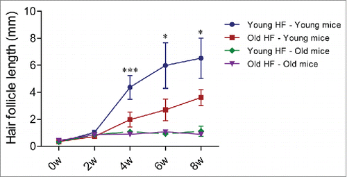
Figure 4. Time-course comparison of ND-GFP fluorescence of HAP stem cells and their location in young and old hair follicles transplanted to young nude host mice. ND-GFP expressing HAP stem cells were located in various areas: sensory nerve, hair matrix bulb area, and outer-root sheath area. HAP stem cell ND-GFP fluorescence was imaged with the Dino-Lite. In the fluorescence images, each follicle is outlined with a dashed line and numbered for comparison with the brightfield images where the follicles are numbered. Please see the Materials and Methods for details.
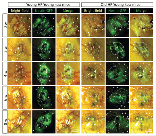
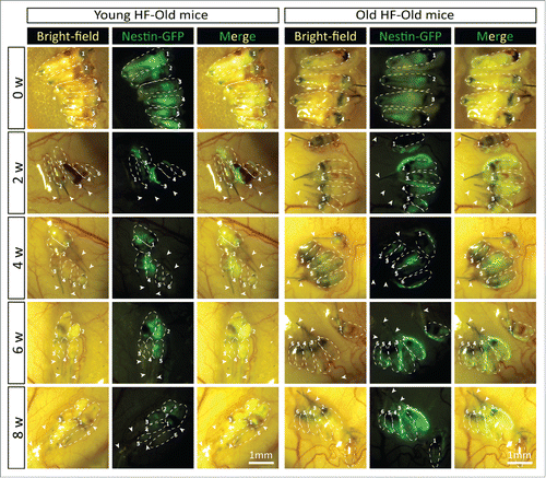
Figure 5. Time-course comparison of ND-GFP fluorescence of HAP stem cells in young and old hair follicles transplanted to old nude host mice. After subcutaneous transplantation. ND-GFP-expressing HAP stem cells in young hair follicles of old nude mice were located mainly in the center of hair follicles (arrows), while in old hair follicles, the ND-GFP expressing HAP stem cells were located mainly in the attached sensory nerves (arrow). HAP stem cell ND-GFP fluorescence was imaged with the Dino-Lite. In the fluorescence images, each follicle is outlined with a dashed line and are also numbered for comparison with the brightfield images where the follicles are numbered. Please see the Materials and Methods for details.