Figures & data
Figure 1. ChIP analysis of Oct-1 at the 5′-CGCCCTGCGTATTCCT-3′ (S100A4) site of the regulatory region of s100a4 gene. The data was quantified and normalized to the level of gene CD3 used as a control. Columns: (1) Namalwa – non-transformed Namalwa cells, (2) NOct1A, (3) NOct1L, (4) NOct1X – the Namalva cells transformed with Oct-1A, Oct-1L, and Oct-1X isoforms respectively. Namalwa – ChIP was performed with anti- Oct-1 antibodies; NOct1A, NOct1L, NOct1X – ChIP was performed with anti-FLAG antibodies. The data corresponds to single biological samples analyzed in triplicates. B. The effect of Oct-1 overexpression on S100A4 transcription level. The relative amounts of mRNA in the transformed cells were quantified and normalized to the corresponding mRNA amounts in the non-transformed cells. The relative transcription level of S100A4 mRNAs in the cells was measured by Real-Time PCR. Error bars show SEM based on 5 biological replicates. A, B *p < 0.05.
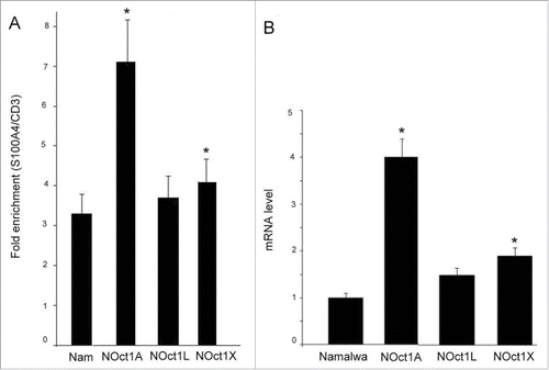
Figure 2. The effect of Oct-1 overexpression on S100A4 protein level in the Namalwa cells (intracellular) and protein secreted by the cells into the culture medium (extracellular). The S100A4 content in the samples was measured by ELISA. NOct1A, NOct1L, NOct1X – Namalwa cells transformed with Oct-1A, Oct-1L, Oct-1X isoforms. Error bars show SEM based on 5 biological replicates. *p < 0.05 B. The effect of Oct-1 overexpression on intracellular S100A4 protein level. Total protein was extracted from the intact Namalwa cells and Namalwa cells transformed with Oct-1A, Oct-1L, Oct-1X isoforms (NOct1A, NOct1L, NOct1X ) and western blotting was performed using antibodies against S100A4. C. Western blot analysis of materials from the Namalwa cells. Intracellular S100A4 protein (cells) and S100A4 secreted into the condition medium (c.m.).
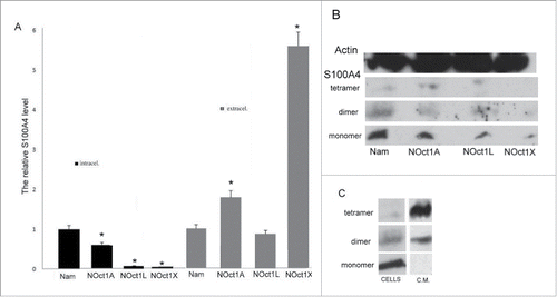
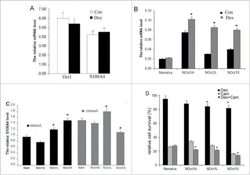
Figure 3. The effect of dexamethasone on Oct-1 and S100A4 transcription levels in the Namalva cells. The relative transcription level of total Oct-1 and S100A4 mRNAs in the cells was measured by Real-Time PCR. B. The resulting effect of dexamethasone and Oct-1-overexpressing on S100A4 transcription level in the Namalva cells. The relative transcription level of total S100A4 mRNAs in the the dexamethasone-treated and in the non-dexamethasone-treated cells was measured by Real-Time PCR. A, B. Error bars show SEM from 5 separate experiments. C. The effect of dexamethasone and Oct-1-overexpressing on S100A4 intracellular level in the Namalva cells (intracellular) and on the secretion of S100A4 by Namalva cells (extracellular). The relative amounts of S100A4 protein in the dexamethasone-treated cells were quantified and normalized to the corresponding amounts of S100A4 protein in non-treated cells. The S100A4 content in the samples was measured by ELISA. The histograms are representative of 5 separate experiments. D. The effect of Oct-1 overexpression on cell viability; the cells treated with dexamethasone or camptothecin or both. Namalwa cell viability was scored by MTT assays. Relative survival was calculated by setting the values in the corresponding non-dexamethasone and non-camptothecin cells as 100%. The histograms are representative of 5 separate experiments. B, C, D *p < 0.05.