Figures & data
Figure 1. Whisker follicles from 4-week-old mice have greater potential for differentiation to cardiac-muscle cells than older mice. (A) Location map of mouse whisker follicles from one pad in one cheek. Blue box = nose side of the mouse whisker pad, Red box = ear side of the mouse whisker pad. (B) Whisker follicles from 4-week-old mice have a greater potential for differentiation to cardiac-muscle cells compared to 10-, 20-, and 40-week-old mice (n = 4 or each time point). (C) Comparison of the differentiation efficiency to cardiac-muscle cells from whisker follicles with mice of different age. (D) Anti-cTnT levels (ELISA analysis) in 4-, 10-, 20- and 40-week-old mouse cardiac-muscle cells differentiated from cultured whiskers. (E) Comparisons of whisker follicles from the ear side and nose side for differentiation potential to cardiac-muscle cells.
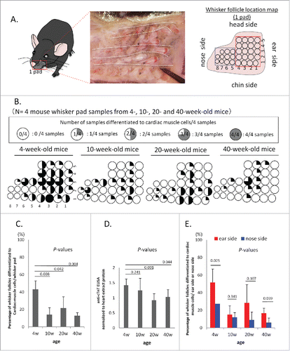
Figure 2. Differentiation to cardiac-muscle cells from whisker follicles from 4-, 10-, 20- and 40-week-old mice. (A) The total differentiated cardiac-muscle cell number from the upper part of whisker follicles from the ear side of the whisker pad, cultured in DMEM with 10% FBS. (B) Number of cTnT-positive cells differentiated from the whisker follicles from the ear side of the whisker pad, shown by flow cytometry. (C) Percentage of cTnT-positive cardiac-muscle cells differentiated from whisker follicles. (D) The number of cardiac-muscle cells differentiated from the upper part of whisker follicles.
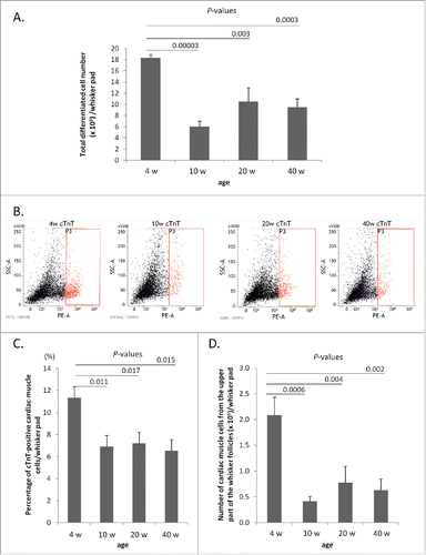
Figure 3. The differentiation potential to various cell types from whisker follicles from mice of different ages. Immunofluorescence staining showed that whisker follicle HAP stemcells from 4-, 10-, 20-, and 40-week-old mice differentiated to cardiac muscle cells, neurons, glial cells, keratinocytes and smooth muscle cells. Red = cTnT, tubulin, GFAP, Keratin 15 (K15) and smooth muscle actin (SMA). Blue = DAPI. Bar = 100 µm. Phase-contrast images showed that HAP stem cell from 4-, 10-, 20-, and 40-week-old mice also differentiated to melanocytes. Bar = 100 µm.
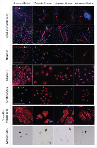
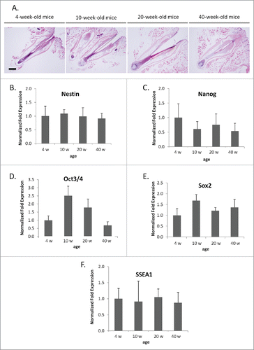
Figure 4. mRNA levels of stem cell marker genes from whisker hair follicles of various age. (A) Hematoxylin and eosin staining of whisker follicles isolated from 4-, 10-, 20-, and 40-week-old mice. Bar = 200 µm. (B-F) Comparisons of mRNA levels of stem-cell-marker genes: nestin (B), Nanog (C), Oct 3/4 (D), Sox 2 (E), SSEA1 (F) were determined with PCR. Results are mean ± SD for 3 samples each. The mRNA level of stem-cell-marker genes did not show any significant differences with whisker age.