Figures & data
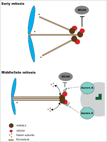
Figure 1. Model for MCRS1 function. At early mitosis MCRS1 (top) interacts with KANSL3 at minus end of microtubules nucleated from chromosomes. This localization at minus ends prevents microtubule depolimeryzation by the action of MCAK and other depolymerases. At later stages in mitosis (bottom), the minus ends of K-fibers are closer to the poles and MCRS1 becomes phosphorylated by Aurora A kinase. This phosphorylation affects MCRS1 activity but not its localization.