907
Views
11
CrossRef citations to date
0
Altmetric
Report

Upregulation of a disintegrin and metalloproteinase-33 by VEGF in human airway smooth muscle cells: Implications for asthma

, , , , , , & show all
Pages 2819-2826 | Received 20 Jul 2016, Accepted 31 Jul 2016, Published online: 31 Aug 2016

Figures & data

Figure 1. VEGF promotes ADAM-33 expression at both mRNA and protein level. ASM cells were incubated with indicated doses of VEGF for 9 h, and then real-time PCR performed. The values are normalized relative to the GAPDH standard (A). ASM cells were incubated at indicated times of VEGF (50 ng/ml), and then real-time PCR performed (B). ASM cells were incubated with indicated doses of VEGF for 24 h (C). ASM cells were incubated at indicated times of VEGF (50 ng/ml), and then western blotting analysis for ADAM-33 was performed. β-actin was used as a loading control (D). All data are representative of 3 independent experiments. Values represent the means ± SEM. *P < 0.05, **P < 0.005 vs. control.
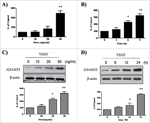
Figure 1. VEGF promotes ADAM-33 expression at both mRNA and protein level. ASM cells were incubated with indicated doses of VEGF for 9 h, and then real-time PCR performed. The values are normalized relative to the GAPDH standard (A). ASM cells were incubated at indicated times of VEGF (50 ng/ml), and then real-time PCR performed (B). ASM cells were incubated with indicated doses of VEGF for 24 h (C). ASM cells were incubated at indicated times of VEGF (50 ng/ml), and then western blotting analysis for ADAM-33 was performed. β-actin was used as a loading control (D). All data are representative of 3 independent experiments. Values represent the means ± SEM. *P < 0.05, **P < 0.005 vs. control.

Figure 2. ADAM-33 siRNA transfection inhibits VEGF-induced cell proliferation. ASM cells were incubated with indicated doses of VEGF for 48 h (A). ASM cells were incubated at indicated times of VEGF (50 ng/ml), and then cell proliferation was determined by BrdU incorporation (B). ASM cells were transfected with negative siRNA or ADAM-33 siRNA, and then real-time PCR performed. The values are normalized relative to the GAPDH standard (C). ASM cells were transfected with negative siRNA or ADAM-33 siRNA, and then western blotting analysis for ADAM-33 was performed. β-actin was used as a loading control (D). ASM cells were transfected with negative siRNA or ADAM-33 siRNA in the presence of VEGF (50 ng/ml) for 48 or 72 h, and then cell proliferation was determined by BrdU incorporation (E). All experiments were done at least twice. Values represent the means ± SEM. *P < 0.05, **P < 0.005 vs. control; # P < 0.05 vs. control siRNA.
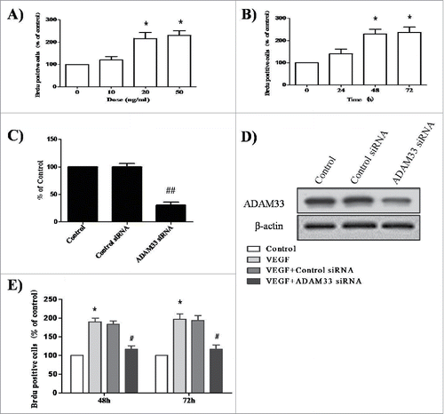
Figure 2. ADAM-33 siRNA transfection inhibits VEGF-induced cell proliferation. ASM cells were incubated with indicated doses of VEGF for 48 h (A). ASM cells were incubated at indicated times of VEGF (50 ng/ml), and then cell proliferation was determined by BrdU incorporation (B). ASM cells were transfected with negative siRNA or ADAM-33 siRNA, and then real-time PCR performed. The values are normalized relative to the GAPDH standard (C). ASM cells were transfected with negative siRNA or ADAM-33 siRNA, and then western blotting analysis for ADAM-33 was performed. β-actin was used as a loading control (D). ASM cells were transfected with negative siRNA or ADAM-33 siRNA in the presence of VEGF (50 ng/ml) for 48 or 72 h, and then cell proliferation was determined by BrdU incorporation (E). All experiments were done at least twice. Values represent the means ± SEM. *P < 0.05, **P < 0.005 vs. control; # P < 0.05 vs. control siRNA.

Figure 3. ADAM-33 siRNA transfection inhibits cell cycle in ASM cells. ASM cells were transfected with negative siRNA or ADAM-33 siRNA in the presence of VEGF (50 ng/ml) for 48 h, and then Flow cytometric analysis for cell cycle was performed. All experiments were done at least twice. Values represent the means ± SEM.
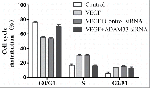
Figure 3. ADAM-33 siRNA transfection inhibits cell cycle in ASM cells. ASM cells were transfected with negative siRNA or ADAM-33 siRNA in the presence of VEGF (50 ng/ml) for 48 h, and then Flow cytometric analysis for cell cycle was performed. All experiments were done at least twice. Values represent the means ± SEM.

Figure 4. Effect of SU1498 on VEGF-induced ADAM-33 expression and cell proliferation in ASM cells. ASM cells were incubated with indicated doses of SU1498 for 2 h before treatment with VEGF (50 ng/ml) for 24 h, and then western blotting analysis for ADAM-33 was performed. β-actin was used as a loading control (A). ASM cells were incubated with indicated doses of SU1498 for 2 h before treatment with VEGF (50 ng/ml) for 48 or 72 h, and then cell proliferation was determined by BrdU incorporation (B). All experiments were done at least twice. Values represent the means ± SEM. *P < 0.05 vs. control; # P < 0.05 vs. VEGF alone.
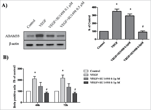
Figure 4. Effect of SU1498 on VEGF-induced ADAM-33 expression and cell proliferation in ASM cells. ASM cells were incubated with indicated doses of SU1498 for 2 h before treatment with VEGF (50 ng/ml) for 24 h, and then western blotting analysis for ADAM-33 was performed. β-actin was used as a loading control (A). ASM cells were incubated with indicated doses of SU1498 for 2 h before treatment with VEGF (50 ng/ml) for 48 or 72 h, and then cell proliferation was determined by BrdU incorporation (B). All experiments were done at least twice. Values represent the means ± SEM. *P < 0.05 vs. control; # P < 0.05 vs. VEGF alone.

Figure 5. Effect of VEGF and SU1498 on phosphorylation of ERK1/2 and Akt in ASM cells. ASM cells were incubated at indicated times of VEGF (50 ng/ml), and then western blotting analysis for phospho-ERK 1/2 (A) and phospho-Akt (B) was performed. ASM cells were incubated with 0.5µM SU1498 for 2 h before treatment with VEGF (50 ng/ml) for 15 min, and then western blotting analysis for phospho-ERK 1/2 was performed (C). ASM cells were incubated with 0.5µM SU1498 for 2 h before treatment with VEGF (50 ng/ml) for 30 min, and then western blotting analysis for phospho-Akt was performed (D). The total ERK1/2 and Akt was used as a loading control. All experiments were done at least twice.
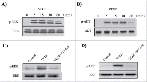
Figure 5. Effect of VEGF and SU1498 on phosphorylation of ERK1/2 and Akt in ASM cells. ASM cells were incubated at indicated times of VEGF (50 ng/ml), and then western blotting analysis for phospho-ERK 1/2 (A) and phospho-Akt (B) was performed. ASM cells were incubated with 0.5µM SU1498 for 2 h before treatment with VEGF (50 ng/ml) for 15 min, and then western blotting analysis for phospho-ERK 1/2 was performed (C). ASM cells were incubated with 0.5µM SU1498 for 2 h before treatment with VEGF (50 ng/ml) for 30 min, and then western blotting analysis for phospho-Akt was performed (D). The total ERK1/2 and Akt was used as a loading control. All experiments were done at least twice.

Figure 6. Effect of LY294002 and U0126 on ADAM-33 expression and cell proliferation in ASM cells. ASM cells were incubated with 20 µM U0126 or 20 µM LY294002 for 2 h before treatment with VEGF (50 ng/ml) for 24 h, and then western blotting analysis for ADAM33 was performed. β-actin was used as a loading control (A). ASM cells were incubated with 20 µM U0126 or 20 µM LY294002 for 2 h before treatment with VEGF (50 ng/ml) for 48 h, and then cell proliferation was determined by BrdU incorporation (B). All experiments were done at least twice. Values represent the means ± SEM. *P < 0.05 vs. control; # P < 0.05 vs. VEGF alone.
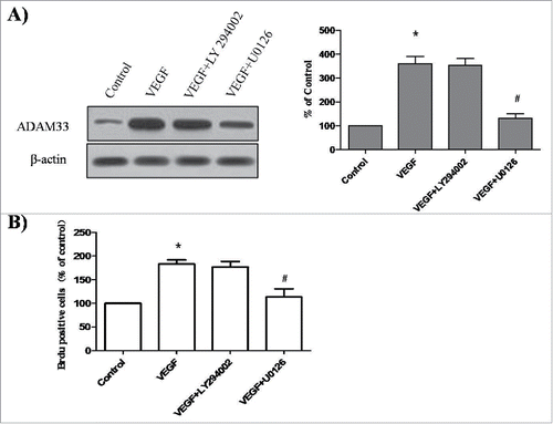
Figure 6. Effect of LY294002 and U0126 on ADAM-33 expression and cell proliferation in ASM cells. ASM cells were incubated with 20 µM U0126 or 20 µM LY294002 for 2 h before treatment with VEGF (50 ng/ml) for 24 h, and then western blotting analysis for ADAM33 was performed. β-actin was used as a loading control (A). ASM cells were incubated with 20 µM U0126 or 20 µM LY294002 for 2 h before treatment with VEGF (50 ng/ml) for 48 h, and then cell proliferation was determined by BrdU incorporation (B). All experiments were done at least twice. Values represent the means ± SEM. *P < 0.05 vs. control; # P < 0.05 vs. VEGF alone.