Figures & data
Figure 1. Klp10A loss leads to overly long centrioles. Centrioles in control (A–C) and mutant (D–F) testes. (A–C) Centrioles elongate gradually in control testes and reach the full length at the end of the first prophase when their distal end organizes a cilium-like projection. Mother (B, arrow) and daughter (B, arrowheads) centrioles have the same length in control germ cells. Mutant centrioles are unusually long with blurred distal ends that often prolonged in single or doublet tubules (D,E, arrowheads); the daughter centrioles (E, arrowhead) are shorter than the mothers (E, arrow) and are displaced from the proximal end of the mothers. Scale bars: 250 nm.
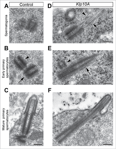
Figure 2. The recruitment of Dplp increases in Klp10A mutant testes. Low magnification of the hub region and early primary spermatocytes in control (A) and mutant (C) testes; centrioles are dot-like in control germline stem cells and early primary spermatocytes, whereas they are very elongated in mutant germ cells (Spd-2, green; Dplp, red; DNA, blue; GSCs, germline stem cells). Representative centrioles in control (B) and mutant (D) male germ cells at indicated stages of development were stained to reveal Spd-2 (green) and Dplp (red). Dplp forms in control testes a short cylinder (B, arrowheads) that elongates in mutant centrioles (D, arrows) and surrounds Spd-2. The paired signals found in mature primary spermatocytes correspond to V-shaped pairs of centrioles. Mother centrioles (D, m) are longer than daughters (D, d) and stained for Spd-2 and Dplp, whereas during the earlier stages of spermatogenesis the daughters often stained only for Spd-2. Acetylated-tubulin labeling (green) reveals distinct cilium-like projections in control mature primary spermatocytes (E, arrowheads) but not during the earlier phases of spermatogenesis in mutant testes; centrioles are stained with Spd-2 (red); GSCs, germline stem cells.Scale bars: A,C = 5 µm; B,D,E,F = 1 µm.
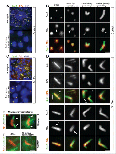
Figure 3. Sas-4 distribution in Klp10A depleted testes. Centrioles in control (A,B) and mutant (C,D) male germ cells at different stages of development were stained to reveal Sas-4 (red), Dplp (green, left column) and Spd-2 (green, right column). (A,B) Dplp and Spd-2 co-localize with Sas-4 during the spermatogonial mitoses although one of the just duplicated centrioles within each pair often lacks Dplp and Spd-2 signals. In mature primary spermatocytes the Sas-4 signal co-localizes with Dplp (A) and is found at the proximal end of the centrioles fully stained by the anti-Spd-2 antibody (B). In Klp10A mutant testes the Sas-4 localization extends over Dplp (C) and Spd-2 (D) signals in 16-cell cyst spermatogonia. (C,D) In mature primary spermatocytes the Sas-4 signal shrinks. Scale bars: 1 µm.
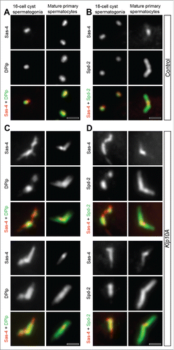
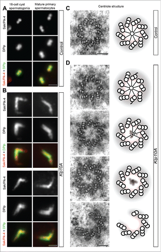
Figure 4. The proximal region of the centriole. Centrioles in control (A) and mutant (B) male germ cells were stained to reveal Sak/Plk4 (red) and Dplp (green). Sak/Plk4 and Dplp co-localize in control testes to the short spermatogonial centrioles or at the proximal end of the giant primary spermatocyte centrioles (A), but expand along the abnormal centrioles in mutant testes (B). Cross sections through the proximal end of control (C) and mutant (D) centrioles: a distinct cartwheel is visible in control centrioles, whereas it is rarely found in mutant centrioles that often lack a cartwheel or display blurred cartwheels. Cartoons depicting the architecture of the basal region of control (C) and mutant (D) centrioles. Scale bars: A,B = 1 µm; C,D = 100 nm.