Figures & data
Figure 1. Isolated human hair follicle and culture of the upper follicle. a. Schema of a human scalp hair follicle shows the location of nestin-positive hHAP stem cells. b. The upper parts of human scalp hair follicles were isolated and cultured in DMEM containing 10% FBS.
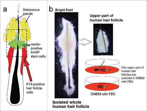
Figure 2. Differentiation of hHAP stem cells. Four weeks after culture in DMEM containing 10% FBS, the upper part of hair follicles differentiated to troponin (cTnT)-positive cardiac-muscle cells, nestin- and βIII-tubulin-positive neurons, GFAP-positive glial cells, K15-positive keratinocytes and smooth-muscle actin (SMA)-positive smooth-muscle cells. Scale bar = 100 µm.
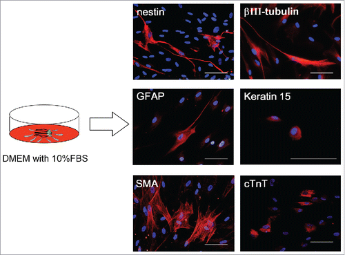
Table 1. FACS analysis of cells differentiated from the upper part of the hair follicle and hHAP stem-cell colonies.
Figure 3. Production of hHAP stem-cell colonies. Human hair follicle culture protocol: isolated upper parts of human hair follicles were cultured in DMEM containing 10% FBS. Four weeks after culture growing cells from the upper parts of human hair follicle were transferred to DMEM/F12 without FBS. One week after culture, the growing cells formed many hHAP stem-cell colonies. Two days after transfer to DMEM containing 10% FBS, hHAP stem-cell colonies started to differentiate.
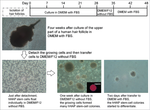
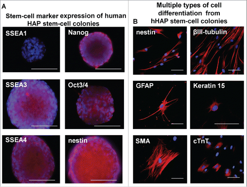
Figure 4. Stem-cell marker expression in hHAP stem-cell colonies and cells differentiated from them. (A) Stem-cell marker expression in hHAP stem-cells colonies. B) Two weeks after transfer to DMEM containing 10% FBS, the nestin-expressing hHAP stem-cell colonies differentiated to troponin (cTnT)-positive cardiac-muscle cells, nestin and βIII-tubulin-positive neurons, GFAP-positive glial cells, K15-positive keratinocytes and smooth-muscle actin (SMA)-positive smooth-muscle cells. Scale bar = 100 μm.