Figures & data
Figure 1. Cell cycle profiles and p27 expression in synchronized 3T3PB and Bcl−2 cells by SS or CI followed by re-stimulation. (A) Bcl−2 expression in 293T, 3T3 and C3H cells. (B, C) Cell cycle profile of 3T3PB and Bcl−2 cells (D, E) Percentage of S phase cells during re-stimulation in serum-starved SS3T3 and contact inhibited 3T3 cells. (F, G). Western blot of p27 in normal growing and synchronized cells. PB: PBABE empty vector stable transfected; Bcl−2: pBABEneo-Bcl−2 stable transfected. CI: contact inhibition SS: serum starvation NG: normal growing
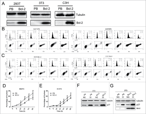
Figure 2. The effects of ATP alteration on cell cycle arrest in 3T3PB and 3T3Bcl−2 cells. (A)The ATP levels during re-stimulation. (B) Cell cycle profiles with addition of various dose of ATP sodium salt after re-stimulation. (C) Graph of the percentage of S phase cells in B. (D) Cell cycle profiles with oligomycinA treatment after re-stimulation. (E) Graph of the percentage of S phase cells in B. (F) Detection of ATP level with oligomycinA treatment.
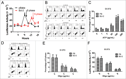
Figure 3. Detection of ROS levels by flow cytometer. (A) ROS levels in synchronized 3T3PB and Bcl−2 cells by CI treatment. (B) ROS levels in synchronized 3T3PB and Bcl−2 cells by SS treatment. (C) Reduction of ROS level by 20 mmol/L GSH treatment.
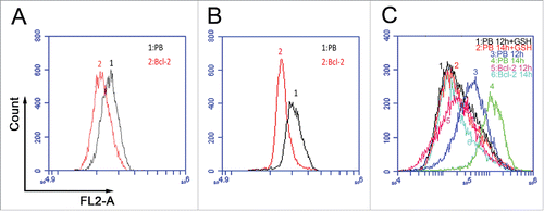
Figure 4. The (S)phase entry profile for PB cells w/o GSH addition during re-stimulation. (A, B) Cell cycle profiles of 3T3PB cells by CI or SS. (C, D) Cell cycle profile of C3HPB cells by CI or SS. (E–F) Graph of percentage of S phase cells in A-D.
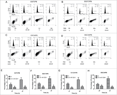
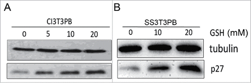
Figure 5. P27 was upregulated by adding 0, 5, 10, 20 mM of GSH.