1,059
Views
23
CrossRef citations to date
0
Altmetric
Report

Combination of gemcitabine and docetaxel regresses both gastric leiomyosarcoma proliferation and invasion in an imageable patient-derived orthotopic xenograft (iPDOX) model

, , , , , , , ORCID Icon, , ORCID Icon, , ORCID Icon & show all
Pages 1063-1069 | Received 27 Jan 2017, Accepted 27 Mar 2017, Published online: 19 May 2017

Figures & data

Figure 1. Imaging of drug efficacy on a leiomyosarcoma iPDOX. The PDOX model treated with GEM combined with DOC (GEM/DOC) demonstrated tumor regression. Scale bar: 5 mm.
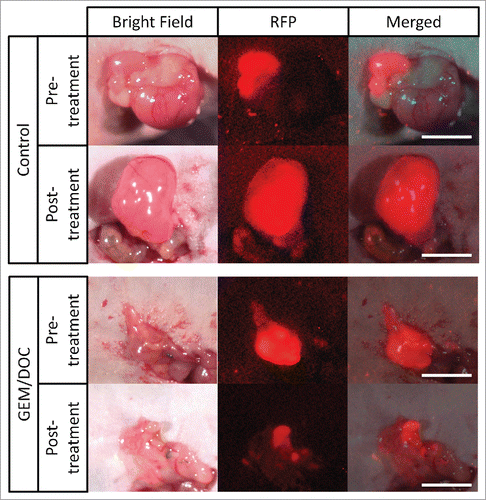
Figure 1. Imaging of drug efficacy on a leiomyosarcoma iPDOX. The PDOX model treated with GEM combined with DOC (GEM/DOC) demonstrated tumor regression. Scale bar: 5 mm.

Figure 2. Efficacy of treatment on leiomyosarcoma iPDOX. Bar graph shows relative tumor volume at a post-treatment point relative to the initial pre-treatment tumor volume. All treatments except for PAZ significantly inhibited tumor growth compared with untreated control (CTR). GEM/DOC was the strongest and significantly more effective than other therapies (DOX: p < 0.01, CPA: p < 0.01, TEM: p < 0.01, YON: p < 0.01, PAZ: p < 0.01). **p < 0.01. Error bars: ± SD.
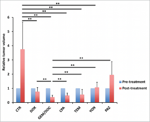
Figure 2. Efficacy of treatment on leiomyosarcoma iPDOX. Bar graph shows relative tumor volume at a post-treatment point relative to the initial pre-treatment tumor volume. All treatments except for PAZ significantly inhibited tumor growth compared with untreated control (CTR). GEM/DOC was the strongest and significantly more effective than other therapies (DOX: p < 0.01, CPA: p < 0.01, TEM: p < 0.01, YON: p < 0.01, PAZ: p < 0.01). **p < 0.01. Error bars: ± SD.

Figure 3. Treatment efficacy for leiomyosarcoma iPDOX. Bar graphs show the tumor fluorescent area (mm2) (A) and fluorescence intensity (B). All treatments significantly inhibited the leiomyosarcoma iPDOX compared with untreated control. **p < 0.01, *p < 0.05. Error bars: ± SD.
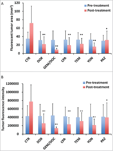
Figure 3. Treatment efficacy for leiomyosarcoma iPDOX. Bar graphs show the tumor fluorescent area (mm2) (A) and fluorescence intensity (B). All treatments significantly inhibited the leiomyosarcoma iPDOX compared with untreated control. **p < 0.01, *p < 0.05. Error bars: ± SD.

Figure 4. Correlation of tumor fluorescent area and tumor fluorescence intensity. Tumor area significantly correlated with fluorescence intensity (R2 = 0.9105, p < 0.0001). Please see Materials and methods for details.
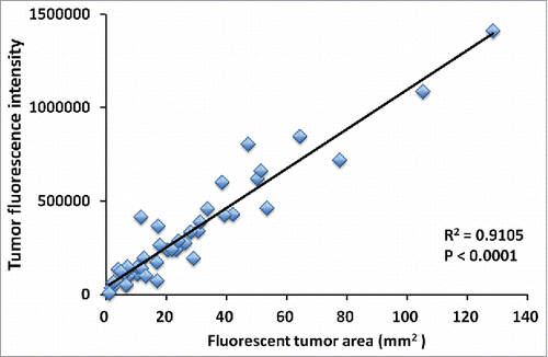
Figure 4. Correlation of tumor fluorescent area and tumor fluorescence intensity. Tumor area significantly correlated with fluorescence intensity (R2 = 0.9105, p < 0.0001). Please see Materials and methods for details.

Figure 5. Correlation of fluorescence intensity and fluorescent area with tumor volume. Both relative tumor fluorescent area (A) and relative fluorescence intensity (B) demonstrated a strong positive correlation with relative tumor volume (R2 = 0.8927, p < 0.0001; R2 = 0.8967, p < 0.0001; respectively).
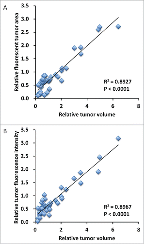
Figure 5. Correlation of fluorescence intensity and fluorescent area with tumor volume. Both relative tumor fluorescent area (A) and relative fluorescence intensity (B) demonstrated a strong positive correlation with relative tumor volume (R2 = 0.8927, p < 0.0001; R2 = 0.8967, p < 0.0001; respectively).
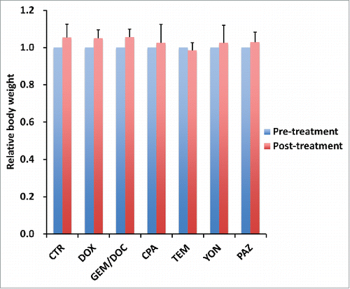
Figure 6. Effect of each treatment on mouse body weight. Bar graph shows relative body weight at post-treatment relative to the pre-treatment. There were no significant differences between each treatment group including untreated control.

Figure 6. Effect of each treatment on mouse body weight. Bar graph shows relative body weight at post-treatment relative to the pre-treatment. There were no significant differences between each treatment group including untreated control.

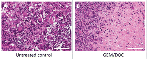
Figure 7. Histological response. A. The untreated control comprised viable cells without necrosis. B. The tumor treated with GEM and DOC exhibited necrosis. Scale bars: 100 μm.

Figure 7. Histological response. A. The untreated control comprised viable cells without necrosis. B. The tumor treated with GEM and DOC exhibited necrosis. Scale bars: 100 μm.

Reprints and Corporate Permissions

Please note: Selecting permissions does not provide access to the full text of the article, please see our help page How do I view content?

To request a reprint or corporate permissions for this article, please click on the relevant link below:

Academic Permissions

Please note: Selecting permissions does not provide access to the full text of the article, please see our help page How do I view content?

Obtain permissions instantly via Rightslink by clicking on the button below:

If you are unable to obtain permissions via Rightslink, please complete and submit this Permissions form. For more information, please visit our Permissions help page.