1,000
Views
34
CrossRef citations to date
0
Altmetric
Report

Salmonella typhimurium A1-R targeting of a chemotherapy-resistant BRAF-V600E melanoma in a patient-derived orthotopic xenograft (PDOX) model is enhanced in combination with either vemurafenib or temozolomide

, , , , , , , ORCID Icon, , , ORCID Icon, , , ORCID Icon & show all
Pages 1288-1294 | Received 15 Feb 2017, Accepted 27 Mar 2017, Published online: 26 Jun 2017

Figures & data

Figure 1. Representative photographs of mice from each treatment group. A. Untreated control. B. Treated with S. typhimurium A1-R. C. Treated with TEM and S. typhimurium A1-R. D. Treated with VEM and S. typhimurium A1-R. Scale bar: 5 mm.
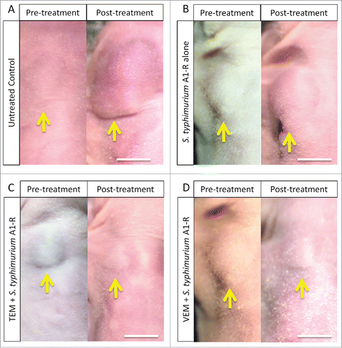
Figure 1. Representative photographs of mice from each treatment group. A. Untreated control. B. Treated with S. typhimurium A1-R. C. Treated with TEM and S. typhimurium A1-R. D. Treated with VEM and S. typhimurium A1-R. Scale bar: 5 mm.

Figure 2. Relative tumor volume in the various treatment groups. Bar graph shows relative tumor volume at post-treatment point relative to the initial pre-treatment tumor volume. Error bars: ± SD.
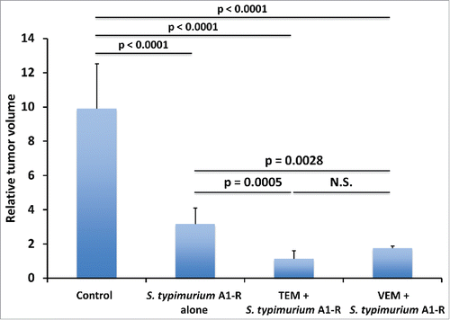
Figure 2. Relative tumor volume in the various treatment groups. Bar graph shows relative tumor volume at post-treatment point relative to the initial pre-treatment tumor volume. Error bars: ± SD.

Figure 3. Fluorescence imaging of S. typhimurium A1-R-GFP targeting alone and in combination with chemotherapy in the melanoma PDOX. Confocal imaging with the FV1000. Scale bars: 12.5 μm.
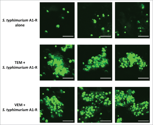
Figure 3. Fluorescence imaging of S. typhimurium A1-R-GFP targeting alone and in combination with chemotherapy in the melanoma PDOX. Confocal imaging with the FV1000. Scale bars: 12.5 μm.

Figure 4. Quantitative tumor targeting by S. typhimurium A1-R-GFP alone and in combination with chemotherapy on the melanoma PDOX model. Bar graphs show S. typhimurium A1-R-GFP fluorescent area (μmCitation2) for each treatment group. Error bars: ± SD.
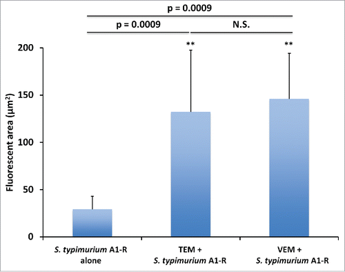
Figure 4. Quantitative tumor targeting by S. typhimurium A1-R-GFP alone and in combination with chemotherapy on the melanoma PDOX model. Bar graphs show S. typhimurium A1-R-GFP fluorescent area (μmCitation2) for each treatment group. Error bars: ± SD.

Figure 5. Histological analysis. Hematoxylin and eosin (H&E) stained slides from tumor tissue of each treatment group. A. Untreated control. B. S. typhimurium A1-R alone. C. S. typhimurium A1-R and TEM. D. S. typhimurium A1-R and VEM. Scale bars: 100 μm.
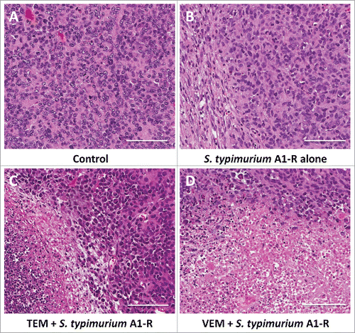
Figure 5. Histological analysis. Hematoxylin and eosin (H&E) stained slides from tumor tissue of each treatment group. A. Untreated control. B. S. typhimurium A1-R alone. C. S. typhimurium A1-R and TEM. D. S. typhimurium A1-R and VEM. Scale bars: 100 μm.

Reprints and Corporate Permissions

Please note: Selecting permissions does not provide access to the full text of the article, please see our help page How do I view content?

To request a reprint or corporate permissions for this article, please click on the relevant link below:

Academic Permissions

Please note: Selecting permissions does not provide access to the full text of the article, please see our help page How do I view content?

Obtain permissions instantly via Rightslink by clicking on the button below:

If you are unable to obtain permissions via Rightslink, please complete and submit this Permissions form. For more information, please visit our Permissions help page.