2,392
Views
58
CrossRef citations to date
0
Altmetric
Extra View

Peroxisomal ACBD4 interacts with VAPB and promotes ER-peroxisome associations

, ORCID Icon, , ORCID Icon & ORCID Icon
Pages 1039-1045 | Received 27 Mar 2017, Accepted 27 Mar 2017, Published online: 05 May 2017

Figures & data

Figure 1 . ACBD4iso2 is a peroxisomal C-tail-anchored protein. (A) Schematic overview of ACBD4iso2 domain structure. ACBD = acyl-CoA binding domain, FFAT-like = 2 phenyalanines in an acidic tract, CC = coiled coil, TMD = transmembrane domain. (B-E) Subcellular localization patterns for ACBD4iso2. COS-7 cells transfected with Myc-ACBD4iso2 were immunolabelled using αPEX14 (peroxisomal marker), αTOM20 (mitochondrial marker) and αMyc antibodies. (E) Higher magnifications of boxed regions are shown (F-G) Differential permeabilisation. COS-7 cells expressing FLAG-ACBD4iso2 were fixed, permeabilised with either Triton X-100 (0.2% in PBS) (F) or digitonin (2.5µg/ml in PBS) (G), and stained with αCatalase (PO matrix), αPEX14 (PO membrane) or αFLAG antibodies. Bars, 10 µm (overlay), 2µm (magnified sections).
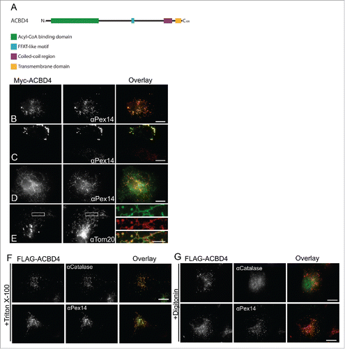
Figure 1 . ACBD4iso2 is a peroxisomal C-tail-anchored protein. (A) Schematic overview of ACBD4iso2 domain structure. ACBD = acyl-CoA binding domain, FFAT-like = 2 phenyalanines in an acidic tract, CC = coiled coil, TMD = transmembrane domain. (B-E) Subcellular localization patterns for ACBD4iso2. COS-7 cells transfected with Myc-ACBD4iso2 were immunolabelled using αPEX14 (peroxisomal marker), αTOM20 (mitochondrial marker) and αMyc antibodies. (E) Higher magnifications of boxed regions are shown (F-G) Differential permeabilisation. COS-7 cells expressing FLAG-ACBD4iso2 were fixed, permeabilised with either Triton X-100 (0.2% in PBS) (F) or digitonin (2.5µg/ml in PBS) (G), and stained with αCatalase (PO matrix), αPEX14 (PO membrane) or αFLAG antibodies. Bars, 10 µm (overlay), 2µm (magnified sections).

Figure 2 . ACBD4iso2 interacts with VAPB. (A) Identification of VAPB and VAPA by MS after co-immunoprecipitation (IP) with GFP-ACBD4iso2 from COS-7 cells (results from 3 experiments); GFP used as control. Only protein IDs which did not appear in any of the GFP only control experiments were considered. (B) Immunoprecipitation (IP) of GFP-ACBD4iso2 and Myc-VAPB after co-expression in COS-7 cells. GFP used as a negative control and GFP-ACBD5 as a positive control. Samples were immunoprecipitated (GFP-Trap) and immunoblotted (IB) using Myc/GFP antibodies.
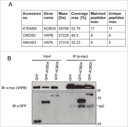
Figure 2 . ACBD4iso2 interacts with VAPB. (A) Identification of VAPB and VAPA by MS after co-immunoprecipitation (IP) with GFP-ACBD4iso2 from COS-7 cells (results from 3 experiments); GFP used as control. Only protein IDs which did not appear in any of the GFP only control experiments were considered. (B) Immunoprecipitation (IP) of GFP-ACBD4iso2 and Myc-VAPB after co-expression in COS-7 cells. GFP used as a negative control and GFP-ACBD5 as a positive control. Samples were immunoprecipitated (GFP-Trap) and immunoblotted (IB) using Myc/GFP antibodies.

Figure 3 . ACBD4iso2/VAPB co-expression promotes PO-ER association. COS-7 cells were transfected with (A) Myc-VAPB alone (immunolabelled using αPEX14, a peroxisomal marker), (B) Myc-VAPB co-expressed with GFP-ACBD4iso2, (C) Myc-VAPB co-expressed with GFP-ACBD4iso2 showing mitochondrial mistargeting. (D) Co-localization of GFP-ACBD4iso2 with Tom20 (mitochondrial marker). Arrows highlight PO-ER association. Bars, 20 µm (overview), 5 µm (cut outs).
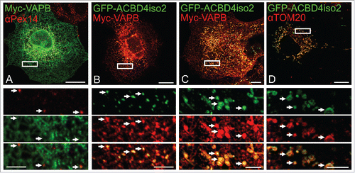
Figure 3 . ACBD4iso2/VAPB co-expression promotes PO-ER association. COS-7 cells were transfected with (A) Myc-VAPB alone (immunolabelled using αPEX14, a peroxisomal marker), (B) Myc-VAPB co-expressed with GFP-ACBD4iso2, (C) Myc-VAPB co-expressed with GFP-ACBD4iso2 showing mitochondrial mistargeting. (D) Co-localization of GFP-ACBD4iso2 with Tom20 (mitochondrial marker). Arrows highlight PO-ER association. Bars, 20 µm (overview), 5 µm (cut outs).

Figure 4 . Model of ACBD4/ACBD5-VAPB interaction. ACBD4 and ACBD5 are both C-tail anchored peroxisomal membrane proteins with functional domains in the cytoplasm which can interact with the MSP domain of ER-resident VAPB via a FFAT-like motif. ACB = acyl-CoA binding, FFAT = 2 phenyalanines in an acidic tract, MSP = major sperm protein binding domain.
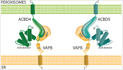
Figure 4 . Model of ACBD4/ACBD5-VAPB interaction. ACBD4 and ACBD5 are both C-tail anchored peroxisomal membrane proteins with functional domains in the cytoplasm which can interact with the MSP domain of ER-resident VAPB via a FFAT-like motif. ACB = acyl-CoA binding, FFAT = 2 phenyalanines in an acidic tract, MSP = major sperm protein binding domain.