Figures & data
Figure 1. β cell specific overexpression of cyclin D2 increases cyclin D2 protein levels. (A) Schematic of the alleles used to create RIP-Cre;cycD2;ROSA26mT/mG mice (KI mice). Black triangles indicate loxP sites. (B) Representative immunofluorescence staining for insulin or dTomato (red), GFP (green), and DAPI (blue) showing efficient Cre recombinase-mediated recombination in KI β cells. (C) Representative immunofluorescence staining for of cyclin D2 (red) and insulin (green) in WT and KI pancreatic sections. (D) Western blot (left panel) and densitometric quantification of cyclin D2 levels (right panel) in isolated islets from WT and KI mice. Data shown as mean ± SD of 3 independent experiments. ** P<0.01, compared with WT mice.
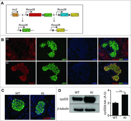
Figure 2. Overexpression of cyclin D2 increases β cell proliferation and β cell mass. (A) Representative immunofluorescence staining for insulin (green), Ki67 (red), and DAPI (blue). White arrows indicate Ins+Ki67+ cells. (B) Quantification of the percentage of β cells expressing Ki67 in WT and KI mice at 3 weeks and 3 months of age. (C) Quantification of β cell mass in WT and KI mice at 3 weeks and 3 months of age. Data shown as mean ± SD (n = 3–4 mice per group). *P < 0.05, **P < 0.01.
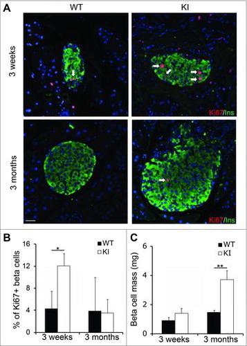
Figure 3. Overexpression of cyclin D2 in β cells does not diminish β cell function. (A) Glucose tolerance test was performed in 3-month-old WT and KI mice. (B) Insulin tolerance was performed in 3-month-old WT and KI mice. (C) Glucose stimulated insulin secretion was measured in 3-month-old WT and KI mice. n = 4–5 animals per group. Data shown as mean ± SD of 3 independent experiments. **P < 0.01.
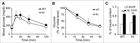
Figure 4. Overexpression of cyclin D2 enhances β cell replication in old KI mice. (A, B) Representative immunofluorescence staining (A) and quantification (B) for insulin (green), Ki67 (red), and DAPI (blue) in young (6-week-old) WT and KI mice challenged with a single dose of STZ (90 mg/kg). C, D. Representative immunofluorescence staining (C) and quantification (D) for insulin (green), Ki67 (red), and DAPI (blue) in old (8-month-old) WT and KI mice challenged with a single dose of STZ (90 mg/kg). White arrows indicate Ins+Ki67+ cells. Data shown as mean ± SD (n = 3–4 mice per group). *P < 0.05, **P < 0.01.
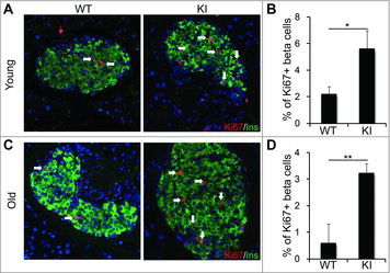
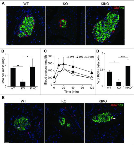
Figure 5. Reconstituting cyclin D2 expression restores β cell mass, function, and regenerative capacity. (A) Representative immunofluorescence staining glucagon (red), insulin (green) and DAPI (blue) in 6-week-old WT, KO, and KIKO mice. (B) Quantification of β cell mass in 6-week-old WT, KO, and KIKO mice. (C) GTT was performed in 3-month-old WT, KO, and KIKO mice. (D, E) Representative immunofluorescence staining (F) and quantification (E) for insulin (green), Ki67 (red), and DAPI (blue) in 6-week-old WT, KO and KIKO mice treated with a single dose of STZ (90 mg/kg). White arrows indicate Ins+Ki67+ cells. Data shown as mean ± SD (n = 3 mice per group). * P < 0.05, **P < 0.01, ***P < 0.005.