Figures & data
Figure 1. Localization of Daam1 during mouse oocyte meiotic maturation. Confocal imaging analysis of Daam1 and actin localization during mouse oocyte meiotic maturation based on staining with an anti-Daam1 antibody. Daam1 co-localized with actin at the cortex of oocyte from the GV stage to the MII stage. Arrow indicates the location of actin cap. Blue: chromatin; Green: Daam1; Red: actin. Bar = 20 μm.
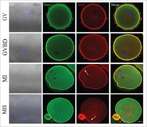
Figure 2. Daam1 knockdown (KD) leads to the failure of mouse oocyte first polar body extrusion. (A) After Daam1 MO injection, Daam1 protein expression was siginficantly reduced by Western blot examination. Band intensity analysis by Image J showed that the intensity of Daam1 significantly decreased after Daam1 knock down. (B) DIC picture showed that oocytes failed to extrude polar body after Daam1 MO injection. Arrows showed that the control oocytes with the polar body while the treated oocytes failed to extrude the polar body. Bar = 100 μm. Analysis of the rate of polar body extrusion showed a significantly decrease after Daam1 knock down. **:significant difference (p < 0 .01).
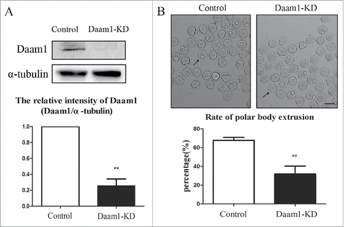
Figure 3. Daam1 affects actin assembly but not spindle position. (A) Actin distribution in oocyte cortex and cytoplasm at MI stage. After Daam1 knock down, the actin distribution was disrupted at both cortex and cytoplasm. Red: actin; Blue: chromatin. Bar = 20 μm. (B) The fluorescence intensity of actin decreased in cortex and cytoplasm after Daam1 knock down. ***: significant difference (p < 0.001). **: significant difference (p < 0.01). (C) For spindle location in the ooctyes, analysis of the rate of spindle to the cortex showed that there was no difference between control groups and treatment groups (p > 0.05). Green, α-tubulin; Blue: chromatin, Bar = 20 μm.
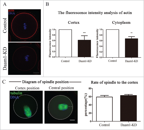
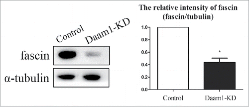
Figure 4. Daam1 knockdown affects the expression of Fascin in mouse oocytes. Fascin protein expression was decreased after Daam1 knock down showing with Western blot analysis. The band intensity of fascin measured by Image J also confirmed this. *: significant difference (p < 0.05).