
Figures & data
Figure 1. HPC transplantation aggravated rat liver fibrosis in the CCl4-induced rat liver fibrosis model. (A) Schematic of the animal experiment (see Methods for details). (B) HE and Sirius Red staining indicated the extent of liver fibrosis, and collagen deposition was examined by Masson's trichrome staining.(C) The expression of α-SMA and CTGF was determined by immunohistochemical staining, brown showed positive expression (n = 5).
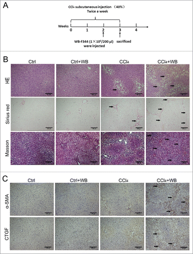
Figure 2. LPS is involved in liver fibrosis in rats and may influence the final fate of HPCs in the CCl4-induced model.(A) Concentration of LPS in portal vein serum was detected using a rat endotoxin ELISA test kit.(B) Schematic of the animal experiment with antibiotic pretreatment (see Methods for details).(C) The level of LPS changed after antibiotic treatment. (D) HE staining indicated the change in liver fibrosis after antibiotic pretreatment.(E) The expression of α-SMA and CTGF was determined by immunohistochemical staining after antibiotic pretreatment.(F) Frozen sections of WB-F344 cells exhibiting green fluorescence in the liver. Data are presented as the mean ± SD. *p < 0.05, **p < 0.01, ***p < 0.001, n = 5.
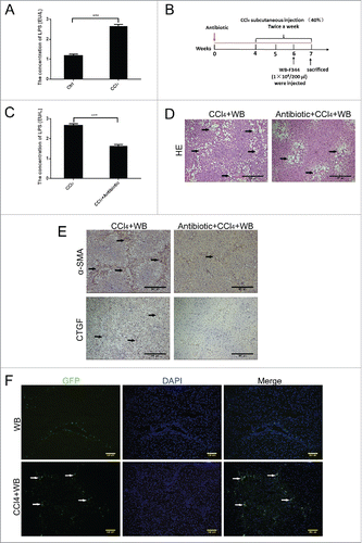
Figure 3. LPS induced the differentiation of WB-F344 cells into myofibroblasts in vitro.(A) Morphological changes of WB-F344 cells in the presence of different concentrations of LPS. (B) qRT-PCR revealed the expression of fibrosis markers (α-SMA, BFGF, CTGF and TGF-β1) in WB-F344 cells treated with LPS for 14 days.(C, D) Western blot analysis and immunofluorescence staining of fibrosis markers (α-SMA and CTGF) in WB-F344 cells treated with LPS for 14 days. Data are presented as the mean ± SD. *p < 0.05, **p < 0.01, ***p < 0.001, NS: no significance.

Figure 4. LPS inhibited the differentiation of WB-F344 cells into hepatobiliary cells in vitro.(A) Morphological changes of WB-F344 cells cultured in hepatocyte differentiation induction medium for 7 days. (B) RT-PCR revealed the expression of hepatocyte markers (Alb, Aldob, cyp1a2 and G6pc) in WB-F344 cells cultured in hepatocyte differentiation induction medium for 7 days.(C) RT-PCR revealed changes in hepatocyte markers in WB-F344 cells cultured with LPS.(D) Morphological changes of WB-F344 cells cultured in bile ductular cell differentiation induction medium for 4 days.(E) RT-PCR revealed the expression of bile ductular cells markers (CK19, GGT and HNF-6) in WB-F344 cells.(F) RT-PCR revealed changes in bile ductular cell markers upon culture with LPS.
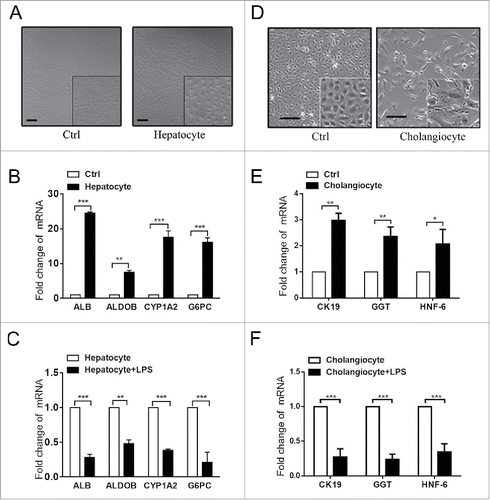
Figure 5. LPS induced the differentiation of WB-F344 cells into myofibroblasts via the Hedgehog signalling pathway.(A) Western blot analysis of key molecules involved in the Hedgehog signalling pathway (Gli-1, Gli-2 and Shh) in WB-F344 cells with or without LPS treatment.(B) Western blot analysis revealed the inhibitory effect of GDC-0449 in WB-F344 cells.(C) Western blot analysis revealed changes in the expression of fibrosis markers (α-SMA and CTGF) in WB-F344 cells after treatment with GDC-0449.
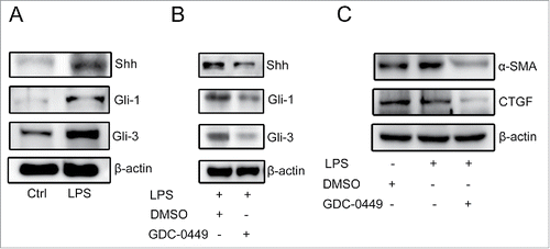
Table 1. Primers used for quantitative real-time RT-PCR.