Figures & data
Figure 1. NC inhibited ovarian cancer cell proliferation and induced apoptosis. (A) Ovarian cancer cell viability was detected after treatment with NC for 24 h, 48 h and 72 h, respectively, using MTT assay. *P < 0.05, **P < 0.01, ***P < 0.001 compared with the control groups treated with DMSO. (B) NC-induced ovarian cancer cell apoptosis was accessed by Flow cytometry.
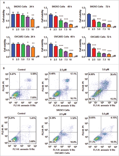
Figure 2. NC induced cell cycke arrest in ovarian cancer cells. (A)NC-triggered cell cycle arrest was measured by Flow cytometry in SKOV3 ovarian cancer cells. (B) Cell cycle analysis was performed in OVCAR3 cells treated with NC.
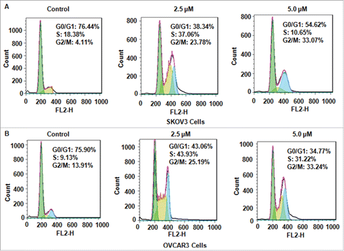
Figure 3. Inhibitory effects of NC on ovarian cancer cell migration and invasion. (A) Left panel: The inhibitory effect of NC on cell migration and invasion was detected using Transwell chambers assay in SKOV3 cells. Right panel: Quantitative results were illustrated for panel A. *P < 0.05, **P < 0.01, ***P < 0.001 compared with the control groups treated with DMSO. (B) Left panel: Cell migration and invasion were measured using Transwell chambers assay in OVCAR3 cells. Right panel: Quantitative results were illustrated for left panel. **P < 0.01, ***P < 0.001 compared with the control groups treated with DMSO. (C) The mRNA expression of Skp2 was measured by real-time RT-PCR in NC-treated ovarian cancer cells. (D) The expression of Skp2, p21 and p27 was detected by Western immunobloting analysis in NC-treated ovarian cancer cells.
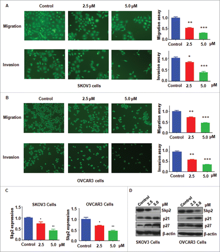
Figure 4. Overexpression of Skp2 abrogated NC-induced cell growth inhibition and cell cycle arrest. (A) The expression of Skp2 was detected by western immunobloting analysis in ovarian cancer cells after Skp2 overexpression plus NC treatment. NC: Nitidine Chloride; NC+cDNA: NC plus Skp2 cDNA transfection. (B) Quantitative results were illustrated for panel A. CTR: Control (C) Ovarian cancer cell growth was measured by MTT assay after Skp2 overexpression in combination with NC treatment. *P < 0.05, vs control; #P < 0.05 vs NC treatment. CTR: Control (D) Cell cycle was accessed by Flow cytometry after Skp2 overexpression in combination with NC treatment.
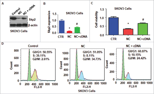
Figure 5. Overexpression of Skp2 abrogated NC-induced apoptosis and invasion inhibition in ovarian cancer cells. (A) Apoptotic cells were accessed by Flow cytometry after Skp2 overexpression in combination with NC treatment. NC: Nitidine Chloride; NC + cDNA: NC plus Skp2 cDNA transfection. (B), Left panel: Cell invasion were detected by Transwell chambers assay in ovarian cancer cells after Skp2 overexpression in combination with NC treatment. Right panel: Quantitative results were illustrated for left panel. **P < 0.01, vs control; #P < 0.05 vs NC treatment.
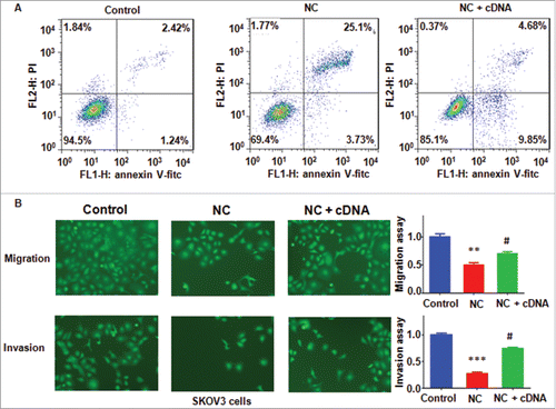
Figure 6. Knockdown of Skp2 enhanced NC-mediated cell proliferation inhibition and cell cycle arrest. (A) The expression of Skp2 was detected by Western blotting analysis in ovarian cancer cells after Skp2 downregulation plus NC treatment. siRNA: Skp2 siRNA; NC+siRNA: NC plus Skp2 siRNA transfection. (B) Quantitative results were illustrated for panel A. CTR: Control. *P < 0.05, vs control; #P < 0.05 vs Skp2 siRNA transfection. (C) MTT assay was performed to detect the effect of Skp2 downregulation in combination with NC treatment on ovarian cancer cell growth. *P < 0.05, vs control; #P < 0.05 vs Skp2 siRNA transfection. (D) Cell cycle was accessed by Flow cytometry after Skp2 downregulation in combination with NC treatment.
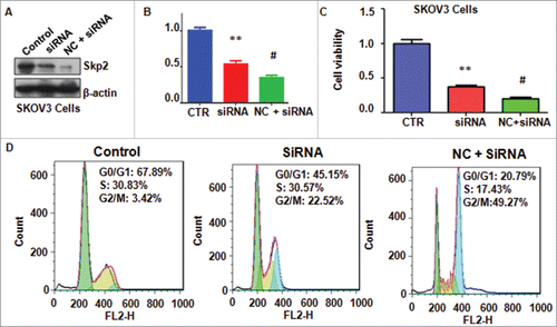
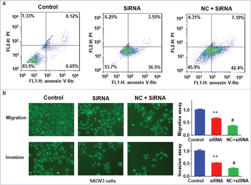
Figure 7. Knockdown of Skp2 enhanced NC-induced cell apoptosis and migration inhibition. (A) Apoptotic cells were detected by Flow cytometry in ovarian cancer cells after Skp2 siRNA transfection and NC treatment. siRNA: Skp2 siRNA; NC+siRNA: NC plus Skp2 siRNA transfection. (B) Left panel: Cell invasion were detected by Transwell chambers assay in ovarian cancer cells after Skp2 siRNA transfection in combination with NC treatment. Right panel: Quantitative results were illustrated for left panel. **P < 0.01, vs control; #P < 0.05 vs Skp2 siRNA treatment.