Figures & data
Figure 1. Overexpression of Skp2 enhanced cell proliferation. (A) The Skp2 mRNA level was detected by real-time PCR in both MG63 and U2OS cells with Skp2 cDNA transfection. (B) Western blot was performed to analyze the protein level of Skp2 in OS cells with Skp2 cDNA transfection. (C) Quantification of the Skp2 protein level was performed. *P < 0.05; ***P < 0.001. (D) Cell viability was measured by CTG solution at 48 hours and 72 hours in MG63 and U2OS cells after Skp2 overexpression.
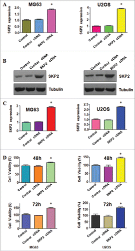
Figure 2. Overexpression of Skp2 inhibited cell apoptosis. (A) Cell apoptosis was determined by Flow cytometry in MG63 cells transfected with Skp2 cDNA. (B) Cell apoptosis was measured by Flow cytometry in U2OS cells with Skp2 overexpression.
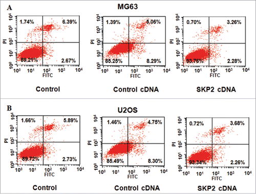
Figure 3. Overexpression of Skp2 enhanced cell cycle progression. (A) The cell cycle analysis was performed in MG63 cells with Skp2 cDNA transfection. X axis: DNA content. (B) The cell cycle analysis was conducted in U2OS cells after Skp2 cDNA transfection. X axis: DNA content.
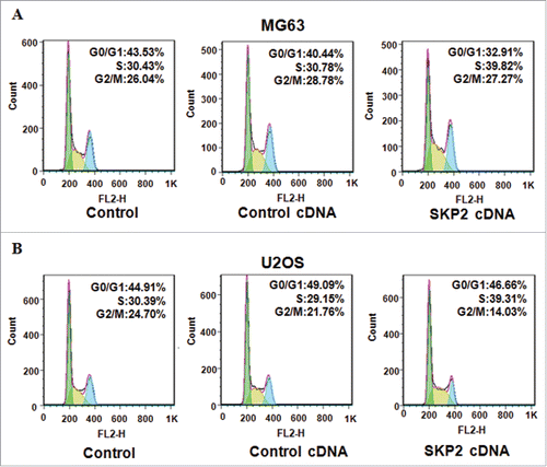
Figure 4. Overexpression of Skp2 promoted cell invasion and migration. (A) Left panel: The ability of cell invasion was measured via transwell assay in OS cells after Skp2 cDNA transfection. Right panel: Quantitative results are illustrated for left panel. (B) Left panel: The ability of cell migration was determined via transwell assay without Matrigel in OS cells after Skp2 cDNA transfection. Right panel: Quantitative results are illustrated for left panel.
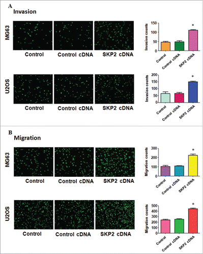
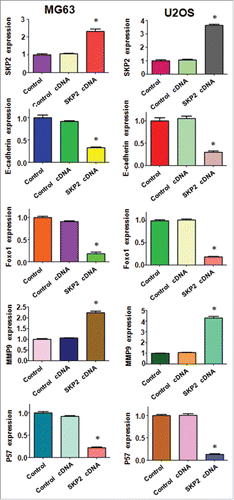
Figure 5. Overexpression of Skp2 regulated the related genes in OS cells. (A) The expression of downstream targets of Skp2 was detected by Western blotting in MG63 cells after Skp2 cDNA transfection. (B) Western blotting was performed to measure the expression of Skp2 downstream targets in U2OS cells with Skp2 overexpression.
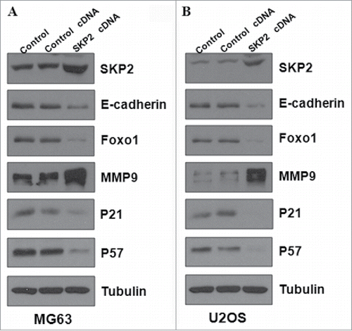
Figure 6. Overexpression of Skp2 governed the downstream genes in OS cells. Quantitative results are illustrated for . ** P < 0.05 vs Control.