1,001
Views
6
CrossRef citations to date
0
Altmetric
Report

Small-molecule CB002 restores p53 pathway signaling and represses colorectal cancer cell growth

, , &
Pages 1719-1725 | Received 09 Jun 2017, Accepted 05 Jul 2017, Published online: 05 Sep 2017

Figures & data

Figure 1. Screening experimental compounds CB002 and a related compound R1 for transcriptional activation by p53 in cancer cells. A. Molecular structure of CB002 and R1. B. SW480 with p53 reporter cells were treated with CB002 and R1 in doses ranging from 0 to 20 µmol/L. p53 responsive bioluminescence was imagined by IVIS. Data are representative of triplicate wells. C. The relative increase of bioluminescence in (B). D. Colorectal cancer SW480, DLD-1, HCT116 and HCT116 p53-null cells were treated with CB002 and R1 for 2 and 24 hour. p53 responsive bioluminescence was imagined by IVIS. E. the relative increased bioluminescence in (D) at 2 hours. F. the relative increased bioluminescence in (D) at 24 hours.
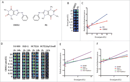
Figure 1. Screening experimental compounds CB002 and a related compound R1 for transcriptional activation by p53 in cancer cells. A. Molecular structure of CB002 and R1. B. SW480 with p53 reporter cells were treated with CB002 and R1 in doses ranging from 0 to 20 µmol/L. p53 responsive bioluminescence was imagined by IVIS. Data are representative of triplicate wells. C. The relative increase of bioluminescence in (B). D. Colorectal cancer SW480, DLD-1, HCT116 and HCT116 p53-null cells were treated with CB002 and R1 for 2 and 24 hour. p53 responsive bioluminescence was imagined by IVIS. E. the relative increased bioluminescence in (D) at 2 hours. F. the relative increased bioluminescence in (D) at 24 hours.

Figure 2. Effect of CB002 and R1 on p53 pathway signaling in cancer cells. DLD-1 and DLD-p73 Knockdown cells (A), HCT116 (B), and HCT116 p53-null cells (C) were treated with CB002 and R1 for 8 and 24 hours. Protein levels of p53 target genes were determined by Western blot analysis.
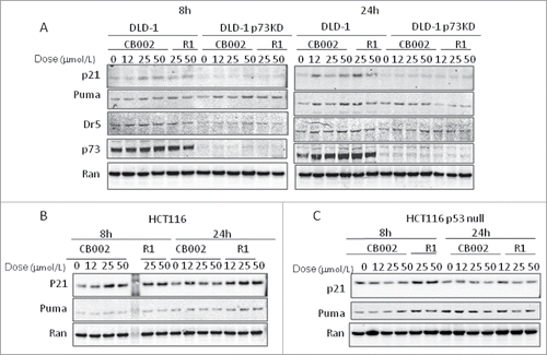
Figure 2. Effect of CB002 and R1 on p53 pathway signaling in cancer cells. DLD-1 and DLD-p73 Knockdown cells (A), HCT116 (B), and HCT116 p53-null cells (C) were treated with CB002 and R1 for 8 and 24 hours. Protein levels of p53 target genes were determined by Western blot analysis.

Figure 3. p53 protein level in cancer cells upon CB002 and R1 treatment. SW480 and HCT116 cells were treated with CB002 and R1 for 24 hours. p53 protein was determined by western blotting using anti-p53 (DO-1).
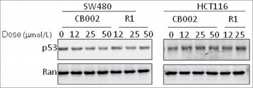
Figure 3. p53 protein level in cancer cells upon CB002 and R1 treatment. SW480 and HCT116 cells were treated with CB002 and R1 for 24 hours. p53 protein was determined by western blotting using anti-p53 (DO-1).

Figure 4. CB002 and R1 induce cell death in colorectal cancer cells. A. Cell viability of SW480 cells treated with CB002 and R1 at 72 hours. B. Cell viability of HCT116 p53-null cells treated with CB002 and R1 at 72 hours. C. Cell viability of HCT116 cells treated with CB002 and R1 at 72 hours. D. Cell cycle profiles of cancer cells SW480, HCT116 and HCT116 p53-null cells. Cells were treated with CB002 and R1 for 72 hrs. Cell viability (A, B, and C) was normalized to DMSO as control. Data are expressed as mean ± SD.
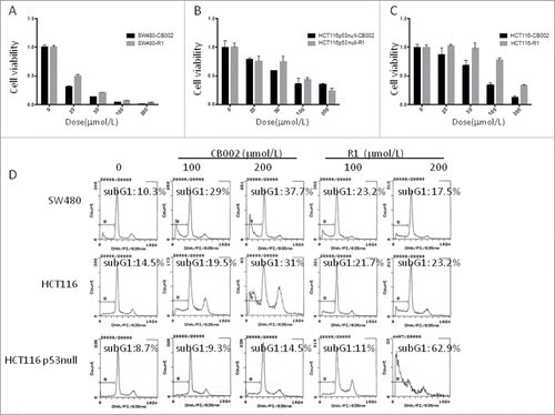
Figure 4. CB002 and R1 induce cell death in colorectal cancer cells. A. Cell viability of SW480 cells treated with CB002 and R1 at 72 hours. B. Cell viability of HCT116 p53-null cells treated with CB002 and R1 at 72 hours. C. Cell viability of HCT116 cells treated with CB002 and R1 at 72 hours. D. Cell cycle profiles of cancer cells SW480, HCT116 and HCT116 p53-null cells. Cells were treated with CB002 and R1 for 72 hrs. Cell viability (A, B, and C) was normalized to DMSO as control. Data are expressed as mean ± SD.

Figure 5. CB002 induces cell death in tumor cells with no significant effect on normal cells. A. Imaging of Cell Titer-Glo as a cell viability assay of SW480 and Wi38 cells treated with CB002 and R1 for 72 hours. B. IC50 of CB002 in cancer cells and normal fibroblast Wi38 cells based on the cell viability. The cells were treated with CB002 for 72 hours. C. Cell cycle profiles of SW480 and Wi38 cells treated with CB002 for 72 hours.
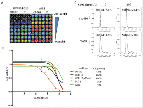
Figure 5. CB002 induces cell death in tumor cells with no significant effect on normal cells. A. Imaging of Cell Titer-Glo as a cell viability assay of SW480 and Wi38 cells treated with CB002 and R1 for 72 hours. B. IC50 of CB002 in cancer cells and normal fibroblast Wi38 cells based on the cell viability. The cells were treated with CB002 for 72 hours. C. Cell cycle profiles of SW480 and Wi38 cells treated with CB002 for 72 hours.

Figure 6. Synergistic effects of CB002 and CPT-11 or 5-FU in treated cancer cells. SW480 cells were treated with CB002 and CPT-11 or 5-FU for 72 hours. A. Imaging of CellTiter-Glo cell viability of SW480 treated with CB002 and CPT-11 or 5-FU. B. Cell viability of SW480 cells treated with CB002 and CPT-11. C. Cell viability of SW480 cells treated with CB002 and 5-FU. D. Combination Index of CB002 and CPT-11. E. Combination Index of CB002 and 5-FU. Cell viability was normalized to DMSO as control. *p < 0.05. Combination index (CI)<1, = 1 and >1 indicate synergism, additive effect and antagonism in drug combination treatment.
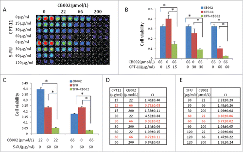
Figure 6. Synergistic effects of CB002 and CPT-11 or 5-FU in treated cancer cells. SW480 cells were treated with CB002 and CPT-11 or 5-FU for 72 hours. A. Imaging of CellTiter-Glo cell viability of SW480 treated with CB002 and CPT-11 or 5-FU. B. Cell viability of SW480 cells treated with CB002 and CPT-11. C. Cell viability of SW480 cells treated with CB002 and 5-FU. D. Combination Index of CB002 and CPT-11. E. Combination Index of CB002 and 5-FU. Cell viability was normalized to DMSO as control. *p < 0.05. Combination index (CI)<1, = 1 and >1 indicate synergism, additive effect and antagonism in drug combination treatment.

Reprints and Corporate Permissions

Please note: Selecting permissions does not provide access to the full text of the article, please see our help page How do I view content?

To request a reprint or corporate permissions for this article, please click on the relevant link below:

Academic Permissions

Please note: Selecting permissions does not provide access to the full text of the article, please see our help page How do I view content?

Obtain permissions instantly via Rightslink by clicking on the button below:

If you are unable to obtain permissions via Rightslink, please complete and submit this Permissions form. For more information, please visit our Permissions help page.