Figures & data
Figure 1. IR up-regulates miR-300 expression in lung cancer cells. (A-D) Human lung cancer cells A549 (A), H446 (B), GLC82 (C) and H1299 (D) were exposed to 2 Gy of X-rays. The relative expression levels of miR-300 were evaluated by qRT-PCR at 0, 6, 24 h post-irradiation. * P < 0.05, ** P < 0.01, compared to control.
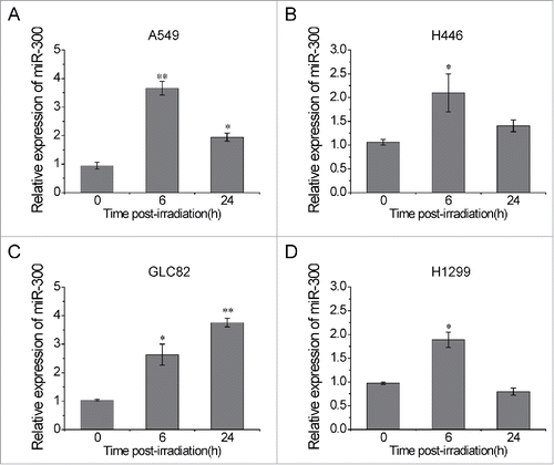
Figure 2. miR-300 restores IR-induced G2 cell cycle arrest. (A) A549 cells transfected with miR-300 mimics or inhibitor were treated with 2 Gy of X-rays, the cell cycle distribution was measured by flow cytometry. Representative images of each group. (B) The quantification of the data in (A). (C-E) Cell cycle distribution of H446 (C), GLC82 (D) and H1299 (E) cells treated with 2 Gy of X-rays at 15 h post-irradiation. (F-G) Cell cycle distribution of A549 (F) and H446 (G) cells treated with 5 Gy of X-rays at 24 h post-irradiation. Ctrl, control; NC, pre-miRNA negative control; P300, pre-miR-300; IN, miRNA inhibitor negative control; I300, miR-300 inhibitor. * P < 0.05, ** P < 0.01, compared to control; NS, not significant.
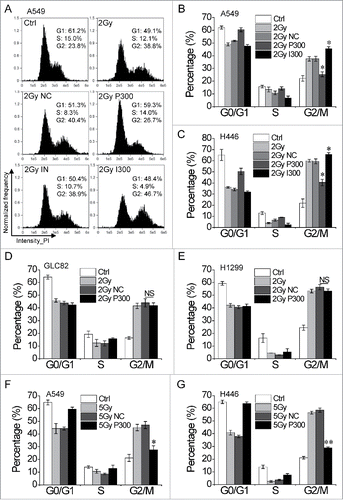
Figure 3. miR-300 targets p53 and apaf1 by binding to mRNA 3′-UTR. (A-B) The sequences of miR-300 and its putative binding sits (rectangle indicated by arrows ) in p53 (A) or apaf1 (B) 3′-UTR. The wild type sequence (WT-P53/APAF1-3′-UTR) or a mutated seed sequence of miR-300-binding site (Mut-P53/APAF1-3′-UTR) were constructed into the luciferase reporter respectively. (C-D) Luciferase reporter containing P53-3′-UTR (C) or APAF1-3′-UTR (D) and miR-300 mimics were co-transfected into A549 cells and the luciferase activity was measured 24 h after transfection. Renilla luciferase activity was used to normalize the firefly luciferase activity. (E) Over-expression of miR-300 down-regulates p53 and apaf1 expression in A549 cells. The levels of p53, p21 and apaf1 were analyzed by western blots 12 h after transfection. (F-H) Over-expression of miR-300 reduces IR-induced p53 and apaf1 expression in A549 (F), H446 (G), H1299 and GLC82 (H) cells. The protein expression levels were measured by western blot 12h after treated with 2 Gy of X-rays. IR, 2 Gy of X-rays irradiation; NC, pre-miRNA negative control; P300, pre-miR-300; +, positive; -, negative. * P < 0.05, compared to NC.
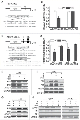
Figure 4. miR-300 modulates radiosensitivity by decreasing p53 and apaf1. (A-B) siRNA targeting p53 (A) or apaf1 (B) interfere its protein expression levels activated by IR. (C-D) miR-300 overexpression or p53 knockdown rescues G2 cell cycle arrest elicited by IR in A549 or H446 cells. Cells transfected with miRNA mimics (NC or P300, 30 nM) or siRNAs (si-NC, or si-p53, 50 nM) were treated with 2 Gy of X-rays The cell cycle distribution was detected 15 h after irradiation by flow cytometry. Representative images of each group in A549 cells (C) and the quantification of each group in H446 cells (D). (E) Survival curves of A549 cells treated with IR after transfected with miR-300 mimics (P300) or p53 siRNA (si-p53). The estimated survival fraction was obtained by fitting to the one-hit multitarget formula at 0, 1, 3 and 5 Gy of X-rays. Data are shown as mean ± SD, the experiment was conducted three times independently. (F) Representative images of colonies in H446 cells treated with 0, 1, 3 and 5 Gy of X-rays. (G) siRNA targeting apaf1 interfere its protein expression levels induced by 5 Gy of X-rays irradiation. The protein levels were analyzed by western blots 12 h after irradiation. (H) The Annexin V-FITC/PtdIns double staining assay in H1299 cells transfected with P300 or si-p53, 24 h following 5 Gy of X-rays irradiation. R, region; PtdIns, propidium iodide; FITC, fluorescein isothiocyanate; IR, 2 Gy of X-rays irradiation; +, positive; -, negative; Ctrl, control; NC, pre-miRNA negative control; P300, pre-miR-300; si-NC, siRNA negative control; si-p53, p53 siRNA; si-apaf1, apaf1 siRNA. * P < 0.05, compared to 2 Gy.
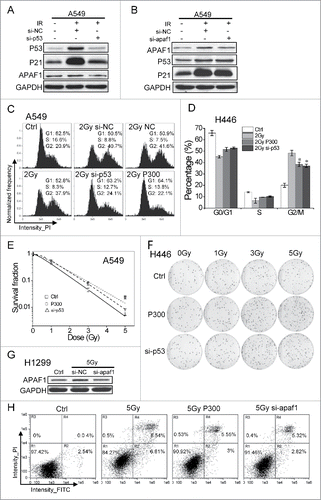
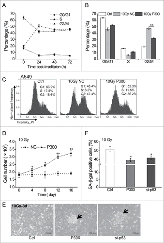
Figure 5. miR-300 reduces p53-dependent senescence induced by IR. (A) High dose irradiation actives cell cycle suspension in A549 cells. A549 cells were exposed to 10 Gy of X-rays, the cell cycle phase was analyzed at 24, 48 and 72 h post-irradiation. (B) miR-300 abolishes cell cycle suspension. The cell cycle phase of A549 cells transfected with miRNA mimics was analyzed at 48 h after irradiation. (C) Representative cell cycle distribution of cells in (B), the data were analyzed with IDEAS Application v6.0. (D) Growth curves of A549 cells treated with 10 Gy of X-rays. Cells were transfected with P300 or NC before irradiation. Graphs represent mean of a triplicate experiment, error bars represent SD. (E) Colonies were formed 8 days after irradiation in miR-300 overexpression (P300) or p53 inhibition (si-p53) group and photographed using a phase contrast microscope. The arrows indicate colonies. Scale bar, 100 μm. (F) Quantitation of SA-β-gal staining A549 cells with miR-300 overexpression (P300) or p53 inhibition (si-p53) 8 days after treated with 10 Gy of X-rays. Ctrl, control, cells exposed to IR but no transfection; NC, pre-miRNA negative control; P300, pre-miR-300; si-p53, p53 siRNA. *P < 0.05, ** P < 0.01, compared to Ctrl.