Figures & data
Table 1. Clinicopathological characteristics
Figure 1. The expression of ALDH1 and CD133 in NSCLC tissues are positively correlated with the clinical features. (A-B) Statistical analysis of ratios of ALDH1+ and CD133+ in NSCLC specimens at different clinical stage using immunofluorescence (IF) and immunohistochemistry (IHC). (C) Representative images of IHC and IF assay of 33 primary NSCLC specimens, including stage I–IV lesions(200 ×). Each bar represents the mean ± SD of three independent experiments. Statistical significance level: p < 0.05.
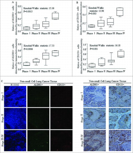
Figure 2. Clinical roles of FBXW7 in patients with NSCLC. (A) The copy number variation of FBXW7 in NSCLC patients, and the genomic alternation is nearly 8% among 119 patients (http://www.cbioportal.org/). (B) The alternation of both mutation and deletion of FBXW7 was referred to shorter disease free survival (http://www.cbioportal.org/). (C-D) Higher level of FBXW7 was correlated with improved overall survival time in about 2000 cases, and means longer survival time before first progression occurred in about 1500 cases (http://kmplot.com/analysis/index.php?p = service&cancer = lung). (E-F) Kaplan-Meier plots results of clinic-pathologic parameter of NSCLC showed no significant differences in cases with or without Let-7c alternations.
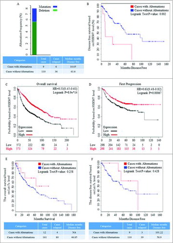
Figure 3. A negative correlation between the expression of FBXW7 and TCF-4 in tissue level. (A) IHC staining for Fbxw7 and TCF4 in tumor-adjacent tissues and NSCLC tissues, original magnification, 200 ×. (B-C) Statistical analysis of Fbxw7 (B) and TCF-4 (C) staining in NSCLC specimens at the different clinical stages (Kruskal-Wallis statistic: 8.464 and14.09, P: 0.0373 and 0.0028, respectively). (D) Using IHC staining, the relative expression and linear correlation of FBXW7 and TCF-4 were analyzed in NSCLC tissues respectively (P < 0.0065, Spearman R: -0.3927).
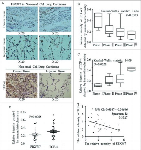
Figure 4. High expression of miR-367 in cancer tissues are closely related to Wnt signaling and let-7c. (A) qRT-PCR analysis of miR-367 and let-7c expression in NSCLC tissues and adjacent non-tumor lung tissues. (P = 0.0015, Student's t-test). (B-C) The relative expression and linear correlation of miR-367 and TCF4 in NSCLC tissues respectively (Spearman R: 0.7286, p < 0.0001, Spearman rank correlation test). (D-E) Analysis of relationship between miR-367 and Wnt1 in NSCLC tissues using the same statistical method (Spearman R: 0.7286, p < 0.0001,). (F-I) Linear regression and qRT-PCR analysis of miR-367 and let-7c expression in tumor tissue (F-G) and adjacent tissue (H-I) respectively (*p < 0.05, both of assay). These experiments were repeated three times, statistical significance level: *p < 0.05.
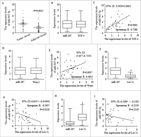
Figure 5. Effect of miR-367, FBXW7 and let-7c on tumor spheroid formation in NSCLC lines. (A-C) Number of spheres per well was quantified on the first generation from A549 (A), H1299 (B), H460 (C) cell lines. (D) Representative images of tumor spheroid form three NSCLC cells with different treatment. (E-G) Number of spheres per well was quantified on the second generation from A549 (E), H1299 (F), H460 (G) cell lines. (H) Representative images of tumor spheroid form three NSCLC cells with different treatment. *P < 0.05 and ** P < 0.01 were based on Student's t-test. ** p < 0.001,* p < 0.01, # p < 0.05.
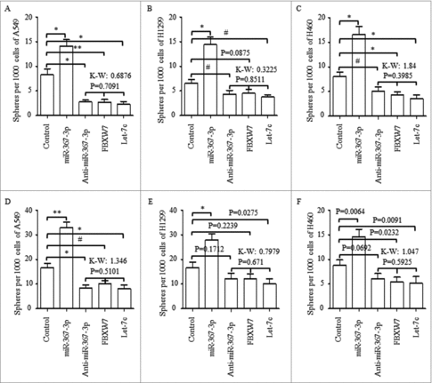
Figure 6. MiR-367 stimulated self-renewal of CSCs via blocking FBXW7 and releasing Let-7c repressed Wnt pathway, A-C. Reintroducing FBXW7, Let-7c or sh-TCF-4 into miR-367 overexpressed spheres cells all abolished its oncogenic roles in self-renewing. TCF-4 inhibition exerted similar effect as the combination of sh-TCF-4 and miR-367 did. D-E. Representative images of spheres from the 1st and 2nd generations were shown. ** p < 0.001,* p < 0.01, # p < 0.05.
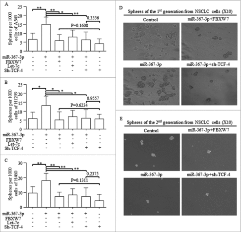
Figure 7. MiR-367 directly targets FBXW7 and represses the level of let-7c through activating Lin28B, (A) The predicted binding sites for miR-367-3p in the 3′-UTR of FBXW7. (B) Luciferase activity was performed in 293 T cells co-transfected with miR-367 mimics or negative control and pGL3 vector containing the wild-type or mutant FBXW7 3′-UTR (p = 0.002, Student's t-test). (C) Dual luciferase activity assays were performed using the promoter reporter plasmid (pGL3-Lin28B-WT or pGL3-Lin28B –Mut) and miR-367 mimics (p = 0.008, Student's t-test). (D) miR-367 significantly repressed the expression of Let-7c in NSCLC cells, p < 0.05 was based on Student's t-test. ** p < 0.001,* p < 0.01, # p < 0.05.
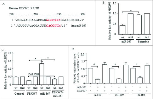
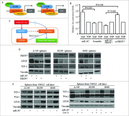
Figure 8. MiR-367 induction of Wnt signaling activation required FBXW7/Let-7c repression. TOP/FOP plasmid was used to detect the FBXW7 controlling of Wnt signaling activity (A), and miR-367 activated Wnt activity effectively (B) No significant difference was detected between groups of miR-367 overexpression and FBXW7 inhibition (B). (C) The hypothesized signaling pathway was drafted and illustrated. (D) Western was used to illustrate the Let-7 dominated pathway, and in lung cancer sphere cells, miR-367 inhibited FBXW7 and consequently released LIN28 and TCF-4 levels. Overexpressing FBXW7 abolished miR-367 stimulation of LIN28 and TCF-4. (E) Factors of Wnt signaling of TCF-4 and Wnt1 increased when miR-367 overexpressed and LIN28 increased, while, reintroducing Let-7c into stem cells attenuated the induction of Wnt signaling by miR-367.