Figures & data
Figure 1. TEAD4 expression is upregulated in CRA. (a) Analysis of TEAD4 expression (RT-PCR, mRNA level) in CRA and their matched adjacent mucosa in Renji dataset. N = 48, paired sample t test. (b) Immunohistochemical analysis of TEAD4 in CRA tissues and paired normal colorectal mucosa (original magnification × 400). (c) Analysis of TEAD4 expression (Immunohistochemical analysis, protein level) in CRA and their matched adjacent mucosa in Renji dataset. N = 48, t test.
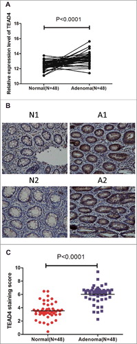
Table 1. The association of TEAD4 expression with clinicopathological characteristics.
Table 2. The association of TEAD4 expression with CRA recurrence.
Figure 2. Roles of TEAD4 in cell proliferation in vitro. (a,b) qPCR and western blot analysis of TEAD4 expression level in the HCT116 and SW480 cells transduced with control siRNA or TEAD4 siRNA. (b) Cell viability of HCT116 and SW480 cells was quantified at the indicated time by using CCK-8 assay. (c) Colony formation assay of HCT116 and SW480 cells transduced with control shRNA or TEAD4 shRNA were performed, stained and examined at 10 days later.
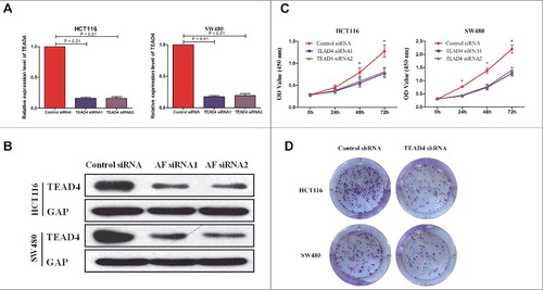
Figure 3. TEAD4 promotes tumor growth in nude mice. (a) Knockdown of TEAD4 inhibits growth of xenografts in nude mice. Stably transfected human CRC HCT116 cells (HCT116/Control shRNA or HCT116/TEAD4 shRNA) were injected subcutaneously into the right forelimb axilla to establish a xenograft model. The sizes of resected tumors arising from cells transfected with/without the TEAD4 knockdown plasmid (at 20 days post-injection) is shown. (b) Tumor growth curves in the TEAD4 knockdown and control groups. (c) Comparison of excised tumor weight in two groups.
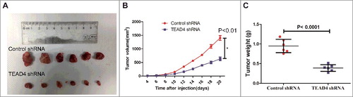
Figure 4. Hippo pathway is regulated by TEAD4 in colon cancer. (a) Heatmap showing the differentially regulated mRNAs in the TEAD4 knockdown HCT116 cells from RNA-seq data analysis. (b) GSEA comparing TEAD4 normal expression group (red) against TEAD4 knockdown expression group (blue) of HCT116 cells. (c) HCT116 and SW480 cells were transfected by TEAD4 siRNA or control siRNA, followed by RT-PCR analysis using specific primers for TEAD4, YAP1 or CTGF. (d) HCT116 and SW480 cells were transfected by TEAD4 siRNA or control siRNA, followed by western blot analysis using specific antibodys for TEAD4, YAP1 or CTGF. (e) Correlation between the mRNA levels of TEAD4 and YAP1 as determined by RT-PCR of CRA tissues. (f) Correlation between the protein levels of TEAD4 and YAP1 as determined by Immunohistochemical staining of CRA tissues.
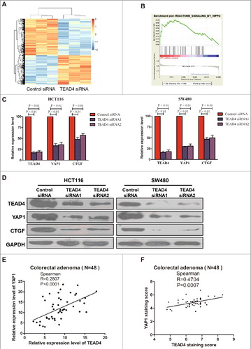
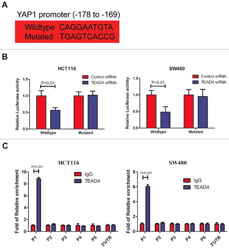
Figure 5. TEAD4 transcriptionally activates YAP1. (a) The predicted TEAD4-binding sequence (−178 to −169) in YAP1 and its mutated variant (both sequences indicated) were respectively inserted into a luciferase reporter and analyzed for their responses to TEAD4. (b) Relative expression of WT and TEAD4-binding motif mutant YAP1 promoter-driven luciferase reporters in HCT116 or SW480 cells transfected with control siRNA or TEAD4 siRNA. (c) ChIP analysis of TEAD4 binding to the YAP1 promoter in the HCT116 or SW480 cells. qPCR was performed with primer specific to regions round the TEAD4-binding motifs (P1) and four control region (P2, P3, P4, P5 and YAP1 3′UTR).