Figures & data
Figure 1. Relocation of S100A4 protein from lymphocytes to the surface of cancer target cell. A,B. Double immunostaining for S100A4 and CD95 revealed exclusive expression of S100A4 protein in lymphocytes (green), while CD95 expression was limited to K562 cell before cell contact (red). С. Immunostaining with anti-S100A4 antibodies detected lymphocytes as well as cancer cell. (10.0x/1.40 NA oil objective). D. Co-localization of S100A4 and CD95 on the surface of K562 target cell after contact with lymphocytes.
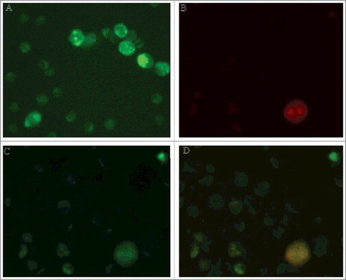
Figure 2. А. Western-blot analysis of CSML-100 surface proteins. В. Western-blot analysis of CSML-100 surface proteins after their interaction with PGRPs. C. Western-blot analysis of recombinant proteins: Hsp70, S100A4, Hsp70 and S100A4 after their interaction with a lymphocyte. А and В. CSML-100 surface proteins were biotinylated; results are visualized using Streptavidin-Horseradish Peroxidase. C. Recombinant Hsp70 and S100A4 are biotinylated; formed complexes are extracted on AbPGRPs-sepharose. D. S100A4 concentration on the CSML-100 cells before and after their stimulation with PGRPs. The S100A4 content in the samples was measured by ELISA. The stimulation was performed by adding 20 ng/ml PGRPs into the CSML-100 incubation medium for 24 hours. Error bars show SEM based on five biological replicates.
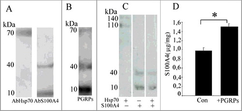
Figure 3. A. S100A4 impact on target cells survival. The activated lymphocytes (20:1) were added to CSML-100 and melanoma M3 cells that were pre-cultivated with S100A4 antibodies and incubated for 3 hours at 37°C. The cells survival was measured with an MTT test in five independent experiments. Error bars are ±SEM. B. The lymphocytes were extracted from the peripheral blood of healthy donors and incubated for 6 d with 1000 U/ml interleukin-2 or with interleukin-2 and 10 μ/ml paclitaxel. Cultivated and not cultivated with paclitaxel lymphocytes were added to К562 cells in the presence of 1 mM recS100A4, and in the presence or absence of 1mM Ca2+. *p < 0.05. Error bars show SEM based on five biological replicates. С. Paclitaxel impact on CSML-100 cells survival in the presence of cultivated and not cultivated with paclitaxel lymphocytes. The part of CSML-100 cells was pre-cultivated with paclitaxel (10 μ/ml), and then cultivated and not cultivated with paclitaxel lymphocytes were added. Error bars show SEM based on five biological replicates, *p < 0.05.
Figure 4. Interaction between CD4+CD25+PGRPs+S100A4+ lymphocytes and target cells in the presence and absence of S100A4 on their surface. А. S100A4 and PGRPs on the lymphocyte's surface form a complex with Hsp70 on the surface of target cell that do not express S100A4 during a lymphocyte-cell contact. B. S100A4 and PGRPs on the lymphocyte's surface do not form complex with Hsp70 on the surface of target cell that express S100A4 during a lymphocyte-cell contact. C. PGRPs on the lymphocyte's surface do not form complex with Hsp70 on the surface of target cell that do not express S100A4 during a lymphocyte-cell contact.