Figures & data
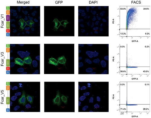
Figure 1. Confocal microscopy (first three columns) and FACS analysis (fourth column) of FcαR variants 1, 3 and 5. HeLa cells were transfected with FcαR variants with C-terminal GFP tags. Variant 1 showed clear fusion protein localization on the membrane whereas variants 3 and 5 showed predominant intracellular localizations. FACS plots of PE-signal on the Y-axis and GFP-signal on the X-axis of HEK293 EXPI cells transfected with GFP fused FcαR variants showed detection of FcαR on extracellular membrane by anti-FcαR-PE antibody only for variant 1.