1,287
Views
22
CrossRef citations to date
0
Altmetric
Research Paper

Adipose-derived stem cell-derived microvesicle-released miR-210 promoted proliferation, migration and invasion of endothelial cells by regulating RUNX3

, , , &
Pages 1026-1033 | Received 10 Nov 2017, Accepted 11 May 2018, Published online: 05 Jul 2018

Figures & data

Figure 1. miR-210 expression in ADSCs-derived microvesicles under hypoxia. ADSCs-derived MVs were divided into two groups: nonhypoxic and hypoxic groups. ADSCs of Hypoxic group were treated with 1% O2 for 24 h, and ADSCs of nonhypoxic group were treated with 20% O2. (a) MV marker (Alix) was measured by western blot. (b) miR-210 expression in ADSCs-derived MVs was measured by qRT-PCR. *P < 0.05, compare with nonhypoxic group.
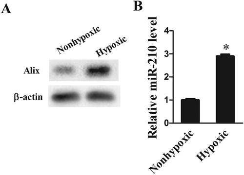
Figure 1. miR-210 expression in ADSCs-derived microvesicles under hypoxia. ADSCs-derived MVs were divided into two groups: nonhypoxic and hypoxic groups. ADSCs of Hypoxic group were treated with 1% O2 for 24 h, and ADSCs of nonhypoxic group were treated with 20% O2. (a) MV marker (Alix) was measured by western blot. (b) miR-210 expression in ADSCs-derived MVs was measured by qRT-PCR. *P < 0.05, compare with nonhypoxic group.

Figure 2. MVs from hypoxic group promoted proliferation and migration of HUVECs. MVs were isolated from ADSCs treated with or without hypoxia, and co-cultured with human umbilical vein endothelial cells (HUVEC). (a) Cell proliferation was detected by MTT assay. (b) Cell migration and invasion were detected by Transwell assay. *P < 0.05, compare with nonhypoxic group.
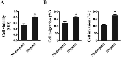
Figure 2. MVs from hypoxic group promoted proliferation and migration of HUVECs. MVs were isolated from ADSCs treated with or without hypoxia, and co-cultured with human umbilical vein endothelial cells (HUVEC). (a) Cell proliferation was detected by MTT assay. (b) Cell migration and invasion were detected by Transwell assay. *P < 0.05, compare with nonhypoxic group.

Figure 3. Effects of miR-210 on the proliferation and migration of HUVECs. miR-210 mimic was transfected into ADSCs under nonhypoxic condition. ADSCs-derived MVs were co-cultured with HUVEC. (a) qRT-PCR showed that miR-210 expression in miR-210 mimic group was upregulated than pre-NC group. (b) Western blot showed that VEGF expression in miR-210 mimic group was upregulated than pre-NC group. (c) MTT assay showed miR-210 mimic promoted proliferation of HUVECs. (d) Transwell assay showed miR-210 mimic promoted migration and invasion of HUVECs. *P < 0.05, compare with pre-NC group. miR-210 inhibitor was transfected into ADSCs under hypoxic condition. ADSCs-derived MVs were co-cultured with HUVEC. (e) qRT-PCR showed that miR-210 expression in miR-210 inhibitor group was downregulated than NC group. (f) Western blot showed that VEGF expression in miR-210 inhibitor group was downregulated than NC group. (g) MTT assay showed miR-210 inhibitor supressed proliferation of HUVECs. (h) Transwell assay showed miR-210 inhibitor supressed migration and invasion of HUVECs. *P < 0.05, compare with NC group.
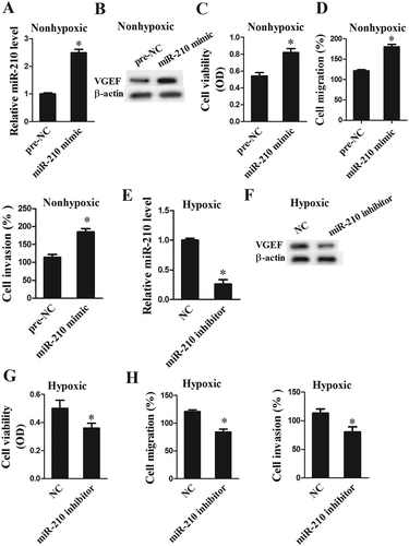
Figure 3. Effects of miR-210 on the proliferation and migration of HUVECs. miR-210 mimic was transfected into ADSCs under nonhypoxic condition. ADSCs-derived MVs were co-cultured with HUVEC. (a) qRT-PCR showed that miR-210 expression in miR-210 mimic group was upregulated than pre-NC group. (b) Western blot showed that VEGF expression in miR-210 mimic group was upregulated than pre-NC group. (c) MTT assay showed miR-210 mimic promoted proliferation of HUVECs. (d) Transwell assay showed miR-210 mimic promoted migration and invasion of HUVECs. *P < 0.05, compare with pre-NC group. miR-210 inhibitor was transfected into ADSCs under hypoxic condition. ADSCs-derived MVs were co-cultured with HUVEC. (e) qRT-PCR showed that miR-210 expression in miR-210 inhibitor group was downregulated than NC group. (f) Western blot showed that VEGF expression in miR-210 inhibitor group was downregulated than NC group. (g) MTT assay showed miR-210 inhibitor supressed proliferation of HUVECs. (h) Transwell assay showed miR-210 inhibitor supressed migration and invasion of HUVECs. *P < 0.05, compare with NC group.

Figure 4. RUNX3 was a direct target of miR-210. (a) Bioinformatics software mircoRNA.org was used to predict microRNAs that bind to RUNX3, and found there were combination sites between miR-210 and 3ʹ UTR of RUNX3. (b) Wide type (WT) and Mutation (MUT) RUNX3 3ʹ UTR were inserted into plasmid pIS0 to construct RUNX3 mRNA (WT) and RUNX3 mRNA (MUT). WT RUNX3-UTR-pIS0 and miR-210 inhibitor were co-transfected into HUVEC. Luciferase activity was higher than NC group, while there was no significant change in luciferase activity after co-transfected with Mu-RUNX3-UTR-pIS0 and miR-210 inhibitor. miR-210 inhibitor was transfected into HUVEC. RUNX3 levels in miR-210 inhibitor group were significantly higher than NC group. (c) WT RUNX3-UTR-pIS0 and miR-210 mimic were co-transfected into HUVEC. Luciferase activity was lower than pre-NC group, while there was no significant change in luciferase activity after co-transfected with Mu-RUNX3-UTR-pIS0 and miR-210 mimic. miR-210 mimic was transfected into HUVEC. RUNX3 levels in miR-210 mimic group were significantly lower than pre-NC group. *P < 0.05, compare with pre-NC or NC group.
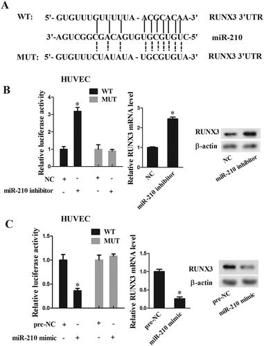
Figure 4. RUNX3 was a direct target of miR-210. (a) Bioinformatics software mircoRNA.org was used to predict microRNAs that bind to RUNX3, and found there were combination sites between miR-210 and 3ʹ UTR of RUNX3. (b) Wide type (WT) and Mutation (MUT) RUNX3 3ʹ UTR were inserted into plasmid pIS0 to construct RUNX3 mRNA (WT) and RUNX3 mRNA (MUT). WT RUNX3-UTR-pIS0 and miR-210 inhibitor were co-transfected into HUVEC. Luciferase activity was higher than NC group, while there was no significant change in luciferase activity after co-transfected with Mu-RUNX3-UTR-pIS0 and miR-210 inhibitor. miR-210 inhibitor was transfected into HUVEC. RUNX3 levels in miR-210 inhibitor group were significantly higher than NC group. (c) WT RUNX3-UTR-pIS0 and miR-210 mimic were co-transfected into HUVEC. Luciferase activity was lower than pre-NC group, while there was no significant change in luciferase activity after co-transfected with Mu-RUNX3-UTR-pIS0 and miR-210 mimic. miR-210 mimic was transfected into HUVEC. RUNX3 levels in miR-210 mimic group were significantly lower than pre-NC group. *P < 0.05, compare with pre-NC or NC group.

Figure 5. miR-210 regulated the proliferation and migration of HUVECs via RUNX3. miR-210 mimic was transfected into ADSCs under nonhypoxic condition, and MVs were isolated. pcDNA-RUNX3 was transfected into HUVEC, then MVs were co-cultured with HUVECs. This experiment included 4 groups: pre-NC, miR-210 mimic, miR-210 mimic+pcDNA, and miR-210 mimic+pcDNA-RUNX3. (a Western blot showed that miR-210 mimic inhibited the expression of RUNX3, and pcDNA-RUNX3 reversed this effect. (b) Western blot showed that miR-210 mimic promoted the expression of VEGF, and pcDNA-RUNX3 reversed this effect. (c) MTT assay showed that miR-210 mimic promoted the proliferation of HUVECs, and pcDNA-RUNX3 reversed this effect. (d) Transwell assay showed that miR-210 mimic promoted the migration of HUVECs, and pcDNA-RUNX3 reversed this effect. miR-210 inhibitor was transfected into ADSCs under hypoxic condition, and MVs were isolated. si-RUNX3 was transfected into HUVEC, then MVs were co-cultured with HUVECs. This experiment included 4 groups: NC, miR-210 inhibitor, miR-210 inhibitor+si-control, and miR-210 inhibitor+si-RUNX3. (e) Western blot showed that miR-210 inhibitor promoted the expression of RUNX3, and si-RUNX3 reversed this effect. (f) Western blot showed that miR-210 inhibitor supressed the expression of VEGF, and si-RUNX3 reversed this effect. (g) MTT assay showed that miR-210 inhibitor supressed the proliferation of HUVECs, and si-RUNX3 reversed this effect. (h) Transwell assay showed that miR-210 inhibitor supressed the migration of HUVECs, and si-RUNX3 reversed this effect. *P < 0.05, compare with pre-NC or NC group. #p < 0.05, compared with miR-210 mimic+pcDNA or miR-210 inhibitor+si-control group.
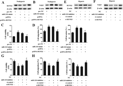
Figure 5. miR-210 regulated the proliferation and migration of HUVECs via RUNX3. miR-210 mimic was transfected into ADSCs under nonhypoxic condition, and MVs were isolated. pcDNA-RUNX3 was transfected into HUVEC, then MVs were co-cultured with HUVECs. This experiment included 4 groups: pre-NC, miR-210 mimic, miR-210 mimic+pcDNA, and miR-210 mimic+pcDNA-RUNX3. (a Western blot showed that miR-210 mimic inhibited the expression of RUNX3, and pcDNA-RUNX3 reversed this effect. (b) Western blot showed that miR-210 mimic promoted the expression of VEGF, and pcDNA-RUNX3 reversed this effect. (c) MTT assay showed that miR-210 mimic promoted the proliferation of HUVECs, and pcDNA-RUNX3 reversed this effect. (d) Transwell assay showed that miR-210 mimic promoted the migration of HUVECs, and pcDNA-RUNX3 reversed this effect. miR-210 inhibitor was transfected into ADSCs under hypoxic condition, and MVs were isolated. si-RUNX3 was transfected into HUVEC, then MVs were co-cultured with HUVECs. This experiment included 4 groups: NC, miR-210 inhibitor, miR-210 inhibitor+si-control, and miR-210 inhibitor+si-RUNX3. (e) Western blot showed that miR-210 inhibitor promoted the expression of RUNX3, and si-RUNX3 reversed this effect. (f) Western blot showed that miR-210 inhibitor supressed the expression of VEGF, and si-RUNX3 reversed this effect. (g) MTT assay showed that miR-210 inhibitor supressed the proliferation of HUVECs, and si-RUNX3 reversed this effect. (h) Transwell assay showed that miR-210 inhibitor supressed the migration of HUVECs, and si-RUNX3 reversed this effect. *P < 0.05, compare with pre-NC or NC group. #p < 0.05, compared with miR-210 mimic+pcDNA or miR-210 inhibitor+si-control group.

Figure 6. MVs-released miR-210 promoted angiogenesis in vivo. Lower-limb ischemia (LI) nude mice model was established. Twenty-five C57Bl/6 mice were divided into four groups: Sham, LI, LI+MVs+NC, and LI+MVs+miR-210 inhibitor groups. (a) Hindlimb muscle tissues were collected 28 days after induction of ischemia, and capillary density was measured. (b) miR-210 expression was detected by qRT-PCR. C. RUNX3 expression was detected by western blot. *P < 0.05, compared with sham group. #p < 0.05, compared with LI+MVs+NC group.
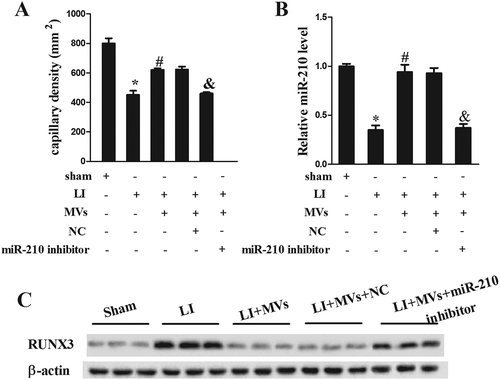
Figure 6. MVs-released miR-210 promoted angiogenesis in vivo. Lower-limb ischemia (LI) nude mice model was established. Twenty-five C57Bl/6 mice were divided into four groups: Sham, LI, LI+MVs+NC, and LI+MVs+miR-210 inhibitor groups. (a) Hindlimb muscle tissues were collected 28 days after induction of ischemia, and capillary density was measured. (b) miR-210 expression was detected by qRT-PCR. C. RUNX3 expression was detected by western blot. *P < 0.05, compared with sham group. #p < 0.05, compared with LI+MVs+NC group.
Supplemental material

Supplemental Material

Download TIFF Image (4.2 MB)

Reprints and Corporate Permissions

Please note: Selecting permissions does not provide access to the full text of the article, please see our help page How do I view content?

To request a reprint or corporate permissions for this article, please click on the relevant link below:

Academic Permissions

Please note: Selecting permissions does not provide access to the full text of the article, please see our help page How do I view content?

Obtain permissions instantly via Rightslink by clicking on the button below:

If you are unable to obtain permissions via Rightslink, please complete and submit this Permissions form. For more information, please visit our Permissions help page.