Figures & data
Table 1. The information of the macaques in this study
Table 2. Primers for the qRT-PCR Assay
Figure 1. BAG3 is increased in astrocytes in the frontal cortex of SHIV-infected macaques
(A-B) Increased BAG3 (brown) in the frontal cortex of SHIV-infected group (#5) (right) compared with control group (#11) (left) was analyzed by IHC. C. Statistical analysis of SHIV-infected group (#1-#8) and control group (#10–13) (***P < 0.001). (D–E) Increased BAG3 in astrocytes of SHIV-infected group (#5) (right) compared with control group (#11) (left) was shown by BAG3 (red) and GFAP (blue) double-labeled IHC. (F) Statistical analysis of SHIV-infected group (#1-#8) and control group (#10–13) (***P < 0.001). (G–H) Western blotting analysis of BAG3 expression in the frontal cortex of SHIV-infected group (#1-#4) compared with control group (#10-#11) and statistical analysis (*P < 0.05). (I) The qRT-PCR analysis of BAG3 mRNA levels in SHIV-infected group (#1-#4) and control group (#10-#11). Statistical analysis of all Western blotting from at least three independent experiments. Ctr: control group; SHIV: SHIV-infected group.
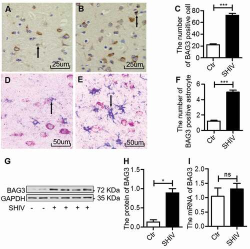
Figure 2. HIV-1 Tat upregulates BAG3 in U87 cells
(A) U87 cells were transfected with pcDNA3.1-Tat-HA (4µg) or pcDNA3.1-HA (4µg) for 48 hours and the expression of HIV-1 Tat was measured by western blotting. (B) U87 cells were transfected with pcDNA3.1-Tat-HA or pcDNA3.1-HA at different time points and the expression of BAG3 was measured by western blotting. (C) Statistical analysis of C (*P < 0.05; **P < 0.01). (D) U87 cells were transfected with pcDNA3.1-Tat-HA or pcDNA3.1-HA at different time points and the mRNA level of BAG3 was measured by qRT-PCR. Statistical analysis of all Western blotting from at least three independent experiments. Tat: pcDNA3.1-Tat-HA; Vehicle: pcDNA3.1-HA.
Figure 3. HIV-1 Tat activates the NF-κB signaling
U87 cells were transfected with pcDNA3.1-Tat-HA only or pcDNA3.1-HA only for 48 hours, or were treated with the NF-κB signaling inhibitor CAPE (30 µg/ml) for 24 hours following transfection with pcDNA3.1-Tat-HA for 24 hours. (A) Expression and nuclear translocation of p65 were observed by confocal microscopy. Red represented expression and distribution of p65, green represented expression of HIV-1 Tat and blue represented nuclear staining by DAPI. All pictures were magnified 600 × . (B) The expression of IκBα, phosphorylated IκBα, phosphorylated p65 and BAG3 were analyzed by western blotting. (C–F) Statistical analysis of B (*P < 0.05; **P < 0.01; ***P < 0.001). Statistical analysis of all Western blotting from at least three independent experiments. Tat: pcDNA3.1-Tat-HA; Vehicle: pcDNA3.1-HA; CAPE: an NF-κB pathway inhibitor.
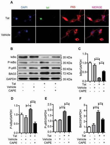
Figure 4. HIV-1 Tat induces autophagy by BAG3-dependent manner
(A) U87 cells were transfected with pEGFP-C3-BAG3-GFP or pEGFP-C3-GFP only for 48 hours, or were treated with the autophagy inhibitor Bafilomycin A1 (Baf-A1) (100 nM) for 24 hours following transfection with pEGFP-C3-BAG3-GFP for 24 hours. The expression of LC3B was analyzed by western blotting. (B) statistical analysis of A (**P < 0.01). (C) U87 cells were transfected with pcDNA3.1-HA or pcDNA3.1-Tat-HA only for 48 hours, or were treated with pcDNA3.1-Tat-HA for 48 hours following transfection with GV112-BAG3-shRNA or GV112-Scramble-shRNA for 24 hours. The expression of BAG3, LC3B and p62 were analyzed by western blotting. (D–F) Statistical analysis of C (**P < 0.01; ***P < 0.001). G. U87 cells were transfected with pcDNA3.1-Tat-HA only or pcDNA3.1-HA only for 48 hours, or were treated with the NF-κB signaling inhibitor CAPE (30 µg/ml) for 24 hours following transfection with pcDNA3.1-Tat-HA for 24 hours. The expression of BAG3, LC3B and p62 were analyzed by western blotting. (H–J) statistical analysis of G (*P < 0.05; **P < 0.01; ***P < 0.001). Statistical analysis of all Western blotting from at least three independent experiments. BAG3: pEGFP-C3-BAG3-GFP; Tat: pcDNA3.1-Tat-HA; Vehicle: pEGFP-C3-GFP or pcDNA3.1-HA; BAG3-shRNA: GV112-BAG3-shRNA; Scramble-shRNA: GV112-Scramble-shRNA; CAPE: an NF-κB signaling inhibitor.