Figures & data
Figure 1. The KEAP1-NRF2 pathway is altered in human lung cancers.
Schematic of KEAP1-NRF2 pathway under homeostatic conditions (left), oxidative conditions (center) or synergistic conditions, for example in a cancer cell (right). Transcriptional activity of NRF2 is highlighted in the green energy bar. Common mutation hotspots in adenocarcinoma (ADC; black circles), squamous cell carcinoma (SqCC; pink circles) or common to both (half black/pink circles) are highlighted in the protein constructs of KEAP1 and NRF2 (NFE2L2). KEAP1, Kelch-like ECH-associated protein 1; NRF2/NFE2L2, Nuclear factor erythoid-2-related factor 2; CUL3, Cullin 3; D, DLG domain; E, ETGE domain; ARE, antioxidant response element; MAF, v-Maf avian musculoaponeurotic fibrosarcoma oncogene homolog; AKT, RAC-alpha serine/threonine-protein kinase; AMPK, AMP-activated protein kinase; GSK3β, Glycogen synthase kinase 3β.
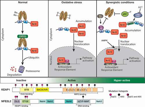
Table 1. Mouse models engineered to harbor KEAP1-NRF2 pathway alterations.
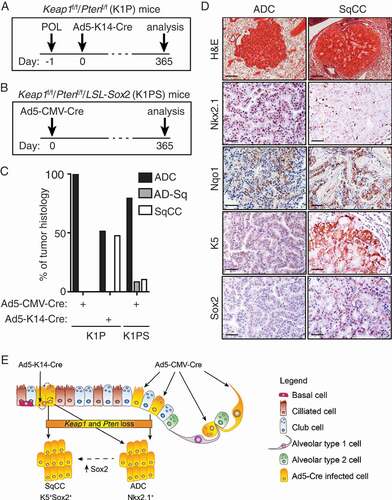
Figure 2. Directed loss of Keap1 and Pten in lung basal progenitor cells promotes lung SqCC formation.
(A) Timeline of polidocanol (POL) administration and adenovirus infection in Keap1f/f/Ptenf/f mice. Briefly, mice were intra-tracheally (i.t.) injected with 10 µl of 2% w/v polidocanol (POL) one day prior to intra-nasal infection of Ad5-K14-Cre virus. Mice were sacrificed, and lung tissue analyzed 12 months following adenoviral infection. (B) Timeline of Ad5-CMV-Cre virus infection in Keap1f/f/Ptenf/f/LSL-Sox2 (K1PS) mice. (C) Quantification of histological phenotype of lung tumors in Ad5-CMV-Cre-infected Keap1f/f/Ptenf/f (K1P) mice [Citation6] (n = 6), Ad5-K14-Cre-infected K1P mice (n = 5) and Ad5-CMV-Cre-infected K1PS mice (n = 3). (D) Representative Hematoxylin & Eosin (H&E) and immunostained sections of lung ADC and SqCC tumors detected in Keap1f/f/Ptenf/f mice 12 months following Ad5-K14-Cre infection. Scale bars; H&E-stained sections 200 µM; Nkx2.1, Nqo1, K5 and Sox2 50 µM. (E) Schematic representation of the consquences of combined loss of Keap1 and Pten in distinct cell types or when combined with enforced Sox2 expression. Combined loss of Keap1 and Pten in bronchiolar and/or alveolar type 2 (AT2) epithelium results in the formation of ADC tumors, while restricted loss of Keap1 and Pten to basal progenitor cells promotes SqCC formation. Interestingly, overexpression of Sox2 in K1P mice promoted the ADC to SqCC transdifferentiation, likely initiated from a “switched” Club or AT2 cell.