Figures & data
Figure 1. AKT signaling pathway is activated by MLN4924 treatment.(a) SK-BR3 and MDA-MB231 cells were incubated with various concentrations of MLN4924 (0, 10, 25 and 50 nM) for 24 h, followed by western blotting using antibodies against p-AKT (S473), t-AKT and NEDD8. ACTIN was used as a loading control. The levels of p-AKT (S473) were quantified and expressed as fold change, compared with the control. (b) SK-BR3 and MDA-MB231 cells were treated with MLN4924 (1 μM) for different time periods (0, 12, 24, 36, and 48 h).
Figure 2. AKT inhibitor MK-2206 enhances the suppression of growth in breast cancer cells by MLN4924.(a) SK-BR3 and MDA-MB231 were treated with indicated concentrations of MK-2206. After 24 h, cells were obtained for western blotting using the indicated antibodies. (b) Cells were treated with the indicated concentrations of MK-2206 for 24 h, 48 h, and 72 h, followed by the ATP-lite assay. (c) Cells were treated with the indicated concentrations of MLN4924 for 72 h and in combination with MK-2206 (100 nM) for 48 h.
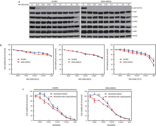
Figure 3. AKT inhibitor MK-2206 enhances the suppression of survival in breast cancer cells by MLN4924.A total of 300 cells were seeded in triplicate in 60-mm dishes and treated with different concentrations of MLN4924 (0, 100, 200 nM) alone or in combination with MK-2206 (100 nM). After 7 days, colonies were stained (a) and counted (> 50 cells in a colony) (b). Rate of colony formation (%) = colony number/300*100% (mean± SEM, n = 3). **p < 0.01.
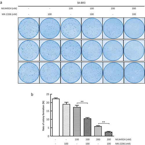
Figure 4. AKT inhibitor MK-2206 enhances the suppression of migration in breast cancer cells by MLN4924.(a) Cells were seeded in 6-well plates and treated with MLN4924 (1 μM) for 24 h, followed by MK-2206 treatment (1 μM) for 24 h. After serum starvation for 12–18 h, the cells were photographed at 0 h, 24 h and 48 h after a pipette tip was used to scratch across the well. Data were shown as the relative wound area normalized to the control (mean± SEM, n = 3). (b) Cells (1x10⁵) were treated with the indicated agents and then seeded into the upper chamber containing serum-free medium. After 24 h, cells were fixed and stained, followed by photography and counting (mean± SEM). (c) Cells were treated with the drugs for 48 h, followed by western blotting using the indicated antibodies. *** p < 0.001.
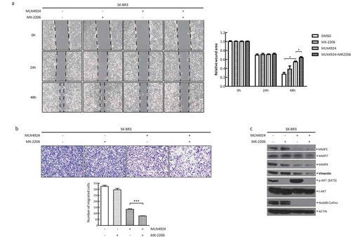
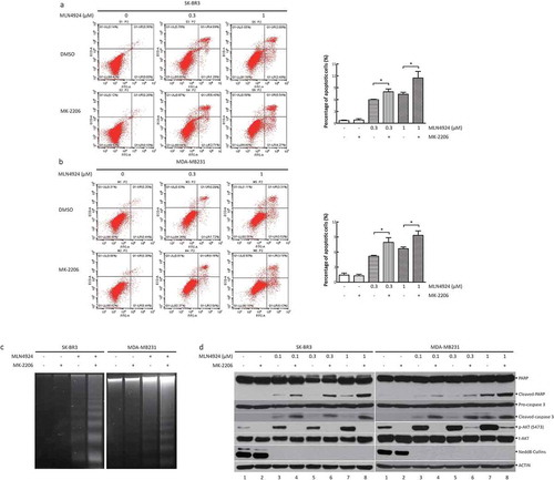
Figure 5. AKT inhibitor MK-2206 enhances the induction of apoptosis induced by MLN4924.(a, b) Cells were treated with MLN4924 (0, 0.3, and 1 μM) alone or in combination with MK-2206 (1 μM). After 48 h, the cells were harvested and stained using the Annexin V-FITC apoptosis detection kit. Cells with Annexin V+ staining located in the right upper and lower quadrants were considered as apoptotic cells (mean± SEM, n = 3). (c) Cells were treated with the drugs for 48 h, followed by DNA extraction. Then, DNA samples were analyzed by agarose gel electrophoresis and were photographed. (d) Cells were incubated with various concentrations of MLN4924 (0, 0.1, 0.3, and 1 μM) for 24 h, followed by MK-2206 (0.5 or 1 μM) or DMSO for 24 h. Cells were harvested for western blotting using indicated antibodies.