Figures & data
Figure 1. clbn knock-out allele has defective S phase checkpoint and intact G2/M checkpoint in response to DNA damage.
The third instar larvae of wild-type fly (w1118) and clbn knock-out mutant were irradiated with 10 Gy. Brains from untreated or irradiated flies were stained with anti-BrdU antibody to detect S phase cells. Representative brains (a-aʹ, b-bʹ) were shown. Quantitative analysis of BrdU labeling cells was shown in (c). Wing discs from w1118 flies (d-dʹ), clbn mutant (e-eʹ) and mei-41 mutant (mei-41 −/-) (f-fʹ) were stained with anti-PH3 antibody to detect mitotic cells. Representative wing discs were shown. Quantitative analysis of mitotic cells was shown in (g). Data was derived from at least five brains or discs for each genotype and three independent experiments. Error bar represents the standard error of mean. *P < 0.001, n.s. not significant.
Figure 2. Clbn expression is induced by irradiation and over-expression of clbn attenuates G1-to-S progression.
The lysate from untreated or irradiated flies were subjected to western-blot analysis. The sample from clbn knock-out mutant was used as a negative control (a). Relative intensity of Clbn western blot band was calculated and shown in (b). The cell cycle profiles of larval brain cells from wild-type w1118 (c), elav-clbn (Cʹ) were detected by flow cytometry. Representative flow cytometry profiles of the cell cycle phases distribution were shown. Quantitative data were presented as mean ± standard error from three experiments. (d–dʹ) Flp-out clones expressing clbn in the brain hemisphere from larval stage, with decreased incorporation of BrdU (green, D) and over-lapping in clbn over-expressing cells (magenta, Dʹ). Flp-out clones expressing clbn in the eye disc (e-eʹ) or wing disc (f-fʹ), with decreased incorporation of EdU (green, E and F) and over-lapping in white dashed circle (Eʹ and Fʹ). MARCM clones of clbn KO in brain lobes, eye disc and wing disc (GFP, Gʹ, Hʹ and Iʹ). Mutant clones lacking clbn exhibited normal BrdU (red, G) and EdU (red, H and I) incorporation. Scale bars, 30 um (D-Dʹ, E-Eʹ, G-Gʹ, H-Hʹ), 10 um (F-Fʹ, I-Iʹ).
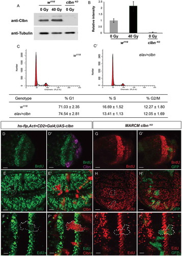
Figure 3. Over-expression of clbn reduces expression of cyclin E and E2F1.
Flp-out clones over-expressing clbn in the wing disc (a-aʹ,c-cʹ,d-dʹ) or eye disc (b-bʹ,e-eʹ,f-fʹ) from larval stage are marked by mRFP (red, (aʹ-fʹ)), stained with anti-cyclin E (green, A and B), anti-cyclin B (green, C and F) and anti-E2F1 antibodies (green, D and E). Clones over-expressing clbn exhibit decreased expression of cyclin E and E2F1, and normal expression of cyclin B. Scale bars, 30 um (a-aʹ,c-cʹ,d-dʹ), 10 um (b-bʹ,e-eʹ,f-fʹ).
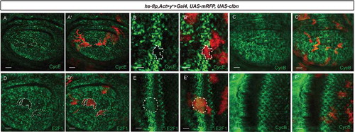
Figure 4. Expression of Clbn is suppressed by E2F1.
(a-aʹ) Wing disc showing expression of Clbn in the posterior region with e2f1 over-expression driven by hh>Gal4. Clbn staining is shown in green, and E2F1 expression is shown in red. Expression of clbn is suppressed by e2f1 over-expression. Flp-out clones over-expressing e2f1 (b-bʹ) or knocking down rbf (c-cʹ) in the wing disc from larval stage are marked by mRFP, and stained with anti-Clbn antibody (green). Clones over-expressing e2f1 or knocking down rbf exhibit decreased expression of Clbn. Wing disc showing expression of Clbn and E2F1 in w1118 (d-dʹʹ). Knockdown of e2f1 (e-eʹʹ,f-fʹʹ) or dp1 (g-gʹ) by hh > Gal4 increases expression level of Clbn. Scale bars, 30 um.
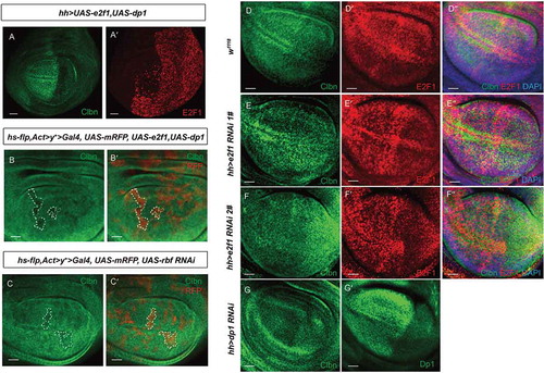
Figure 5. Clbn antagonizes E2F1 activity to regulate cyclin E expression and DNA replication.
Flp-out clones over-expressing e2f1 in the wing disc (a-aʹ,c-cʹ) or eye disc (b-bʹ) from larval stage are marked by mRFP, and stained with anti-cyclin E antibody (green, a-aʹ,b-bʹ) and EdU (green, c-cʹ). Clones over-expressing e2f1 exhibit elevated expression of cyclin E and incorporated EdU, and clbn over-expression simultaneously suppresses the induced expression of cyclin E (d-dʹ and e-eʹ) and incorporated EdU (f-fʹ). Scale bars, 30 um.
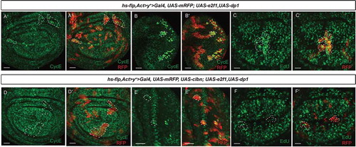
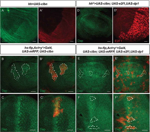
Figure 6. Dap expression is reversely regulated by Clbn and E2F1.
(a-aʹ) Over-expression of clbn driven by hh > Gal4 increases expression of Dap in the posterior compartment. Flp-out clones over-expressing clbn in the wing disc (b-bʹ) or eye disc (c-cʹ) from larval stage are marked by mRFP, and stained with anti-Dap antibody (green). Clones over-expressing clbn exhibit elevated expression of Dap. (d-dʹ) Co-expression of clbn and e2f1 driven by hh > Gal4 decreases expression of Dap in the posterior compartment caused by clbn over-expression. Flp-out clones co-expressing clbn and e2f1 in the wing disc (e-eʹ) or eye disc (f-fʹ) from larval stage are marked by mRFP, and stained with anti-Dap antibody (green). Over-expressing e2f1 reduced the enhanced expression of Dap driven by clbn over-expression. Scale bars, 30 um.