Figures & data
Table 1. Primer sequences.
Figure 1. A successful mouse model of fungal keratitis is established. (a), Comparison of corneal state after the 7th day of Candida albicans infection in mice; (b), Clinical scoring in the Candida albicans group and the saline group; (c), The HE staining of the corneal tissue in mice at the 7th day after modeling with black arrows indicating corneal stromal fibers and red arrows indicating inflammatory cells (× 400), *, p < 0.05; HE, hematoxylin-eosin; n = 12 in the saline group and n = 10 in the Candida albicans group; all data were measurement data, expressed as mean ± standard deviation and analyzed by t-test; the experiment was repeated three times.
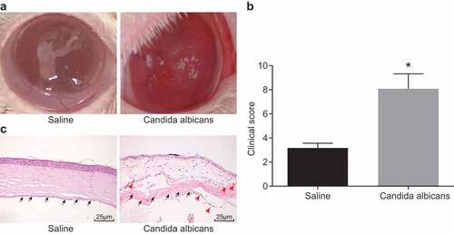
Figure 2. Mice with fungal keratitis present with increased inflammatory cytokine secretion and inflammatory cell infiltration. (a), The images of macrophages (F4/80 positive), neutrophile granulocytes (Ly6g positive) and Th17 (RORγ positive) in the Candida albicans group and the saline group (× 200); (b), The histogram of the positive cell rate of three different cell types; (c), The histogram of relative mRNA levels of different cytokines in each group; (d), The histogram of protein levels of different cytokines in each group; TNF, tumor necrosis factor; IL, interleukin; *, p < 0.05 vs. saline group; n = 12 in the saline group and n = 10 in the Candida albicans group; all data were measurement data, expressed as mean ± standard deviation and analyzed by t-test; the experiment was repeated for three times.
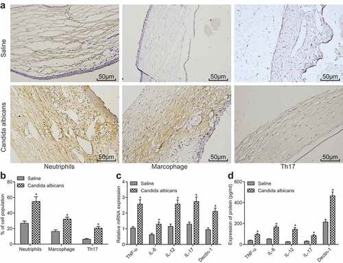
Figure 3. IL-17 gene silencing in Th17 cells inhibits activation of vascular endothelial cells. (a), The histogram of IL-17 level in Th17 (siRNA-NC) and Th17 (siRNA-IL-17) groups; (b), Relative mRNA levels of VCAM-1, E-selectin and Col-IV in Th17 (siRNA-NC) and Th17 (siRNA-IL-17) groups; (c), Relative mRNA levels of VCAM-1, E-selectin, Col-IV and CX43 in the Candida albicans and saline groups; (d), The histogram of mRNA levels of CX43 in different conditions; (e), The images of CX43 expression detected by immunofluorescence (× 200); IL, interleukin; Col-IV, Collagen 4; VCAM-1, vascular cell adhesion molecule 1; CX43, connexin43; *, p < 0.05; n = 12 in the saline group and n = 10 in the Candida albicans group; all data were measurement data, expressed as mean ± standard deviation and analyzed by t-test; the experiment was repeated for three times.
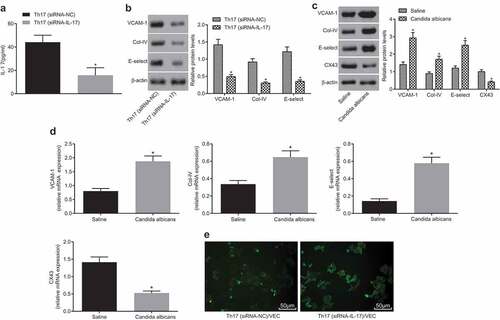
Figure 4. The activation of vascular endothelial cells is inhibited by overexpressing CX43. (a), Relative protein level of CX43 in NC and CX43 groups; *, p < 0.05 vs. the NC group; (b), Relative protein level of CX43 in saline group, IL-17 (1 μM) and IL-17 (5 μM); *, p < 0.05 vs. the blank group; #, p < 0.05 vs. IL-17 (1 μM); (c), Relative protein levels of VCAM-1, E-selectin and Col-IV in each group; (d), Relative mRNA levels of VCAM-1, E-selectin and Col-IV in each group; (c) and (d), * p < 0.05 vs. the blank group; &, p < 0.05 vs. the IL-17 group; NC, negative control; IL, interleukin; CX43, connexin43; all data were measurement data, expressed as mean ± standard deviation; data in panel A were analyzed by t-test, and the other data were tested by one-way analysis of variance; the experiment was repeated for three times.
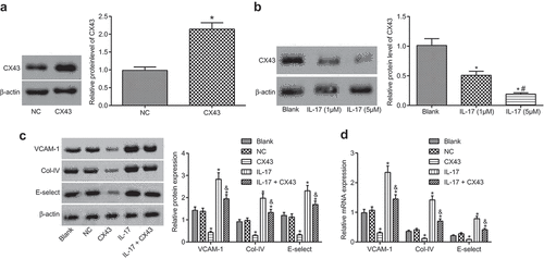
Figure 5. The symptoms of fungal keratitis are alleviated after CX43 knockdown. (a), The expression of CX43 in mouse vascular endothelial cells determined by RT-qPCR; (b), Comparison of clinical scoring in each group; (c), Mice morphological characteristics of corneal tissues in three groups (× 400) with black arrows indicating corneal stromal fibers and red arrows indicating inflammatory cells; (d), Fungal load capacity in infected cornea of each group; (e), Relative mRNA level of each gene in corneal tissues of each group; (f), Protein levels of each gene in corneal tissues of each group; *, p < 0.05 vs. the WT group; #, p < 0.05 vs. the WT + Candida albicans group; HE, hematoxylin-eosin; TNF, tumor necrosis factor; CX43, connexin43; IL, interleukin; n = 12; all data were measurement data, expressed as mean ± standard deviation and analyzed by one-way analysis of variance; the experiment was repeated for three times.
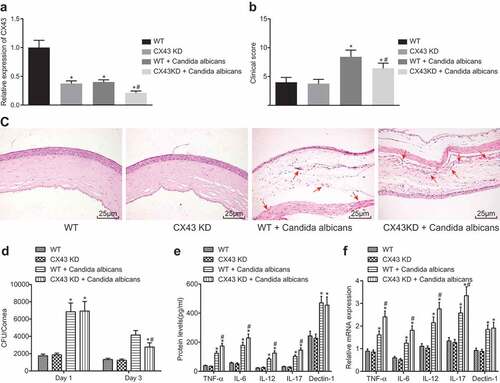
Figure 6. IL-17 regulates endothelial cell activation through the AKT signaling pathway. (a), The gray values of PI3K, p-PI3K, p-AKT, AKT, E-selectin, VCAM-1, COL-IV and CX43 protein bands; (b): The histogram of relative protein levels of PI3K, AKT, E-selectin, VCAM-1, COL-IV and CX43 as well as the extent of AKT and PI3K phosphorylation; *, p < 0.05 vs. the blank group; #, p < 0.05 vs. the IL-17 group; IL, interleukin; Col-IV, Collagen 4; VCAM-1, vascular cell adhesion molecule 1; AKT, serine/threonine kinase; all data were measurement data, expressed as mean ± standard deviation and analyzed by one-way analysis of variance; the experiment was repeated for three times.
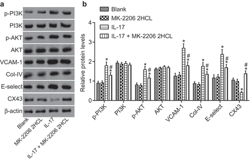
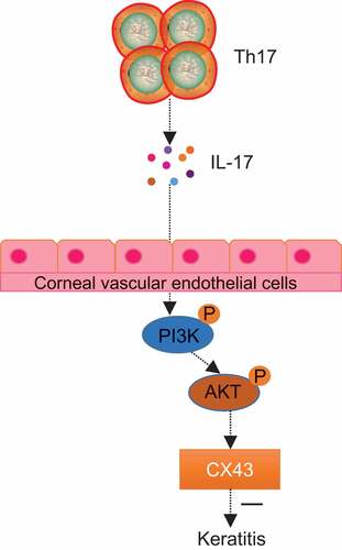
Figure 7. IL-17 inhibited fungal keratitis by reducing the level of CX43 through the AKT signaling pathway. In fungal keratitis, IL-17 secreted by Th17 cells could inhibit the expression of CX43 in the limbal vascular endothelial cells through the AKT signaling pathway, thus inhibiting the development of fungal keratitis.