Figures & data
Figure 1. Fbxo6 is phosphorylated during mitosis. (a-d) HEK293 cells were transfected with flag-tagged Fbxo6 (a), Fbxo4 (b), Fbxo22 (c) or Fbxo36 (d) for 24 hours, respectively. Then the cells were treated with nocodazole (200 ng/ml) for 16 hours (Add for addition) and released for 8 hours (Rel for release). The cells were collected and equal amounts of cell lysates were blotted for anti-flag antibody. (e) HeLaflag-Fbxo6 cells were treated with or without nocodazole (200 ng/ml) for 16 hours. Cell lysates were collected in the presence or absence of lambda protein phosphatase (λ-ppase) and the indicated proteins were examined by western blot. (f) HeLaflag-Fbxo6 cells were treated with nocodazole (200 ng/ml) for 16 hours, and then with or without MG132 (20 μM) for another 3 hours. At the meantime, cells were treated with Roscovitine (20 μM) or Volasertib (50 nM) for 3 hours as well, and the indicated proteins were examined by western blot.
Figure 2. Phosphorylation of Fbxo6 is dynamically changing in cell cycle. (a) HeLa cells stably transfected with flag-Fbxo6 (HeLaflag-Fbxo6) were treated with thymidine (Thy) (2.5 mM) for 24 hours followed by nocodazole (200 ng/ml) treatment for the various times as indicated. DNA content of the treated cells was analyzed by flow cytometry (Asy for asynchronized HeLaflag-Fbxo6cells). (b)The indicated proteins were examined by western blot. (c) HeLaflag-Fbxo6 cells were treated with nocodazole (Noc, 200 ng/ml) for 16 hours and then released for the various times as indicated. DNA content of the treated cells was analyzed by flow cytometry. (d) The indicated proteins were examined by western blot.
Figure 3. Overexpression of Fbxo6 induces premature sister-chromatid separation. (a) The proliferation rate of HeLacon and HeLaflag-Fbxo6 cells for 5 days were analyzed by CCK8 assay. (b) DNA content of HeLacon and HeLaflag-Fbxo6 cells for 3 days was analyzed by flow cytometry. (c) HeLacon and HeLaflag-Fbxo6 cells were treated with nocodazole (200 ng/ml) for 16 hours. Mitotic chromosome spreads were prepared and stained by Wright-Giemsa dye. Arrows indicate prematurely separated sister chromatids. Representative images were shown. The percentages of cells with premature sister-chromatid separation were analyzed. P = 0.0004 vs, control group.
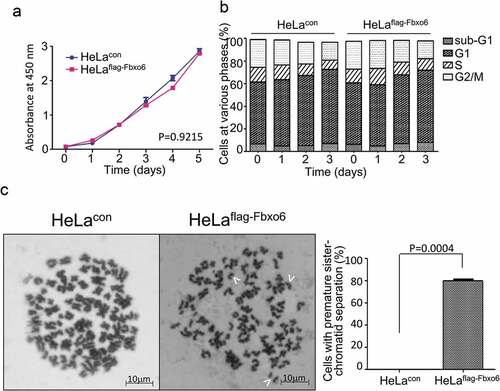
Figure 4. Overexpression of Fbxo6 accelerates the exit from mitosis in nocodazole-released cells. HeLacon and HeLaflag-Fbxo6 cells were treated with nocodazole (200 ng/ml) and then released for the various times as indicated. (a) DNA content of the treated cells was analyzed by flow cytometry. (b) Cells treated in (a) were stained by Wright-Giemsa dye. Representative images were shown. (c) Percentages of cells with binuclear and multi-lobed nuclei, as well as dead cells were counted from cells treated in (a).
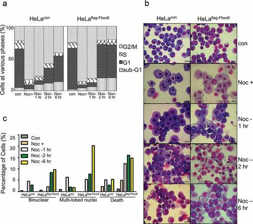
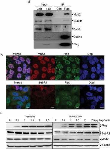
Figure 5. Fbxo6 interacts with spindle check point proteins. (a) Lysates from HeLacon and HeLaflag-Fbxo6 cells stably expressing flag-con or flag-Fbxo6 were immunoprecipitated with anti-FLAG M2 affinity gel. Bound proteins were eluted with FLAG peptide and subjected to Western blot with BubR1, Mad2, Bub3, Cullin1 and flag antibodies. (b) HeLaflag-Fbxo6 cells were stained with Mad2 or BubR1 (Red), Fbxo6 (Green) and Dapi (Blue). (c) HeLa cells were transfected with flag-Fbxo6 at different dosages, and also arrested by thymidine (2.5 mM) at G1/S phase or by nocodazole (200 ng/ml) at G2/M phase. The indicated proteins were examined by western blot.