Figures & data
Figure 1. Pink1 is not present in mitochondria fraction from skeletal muscle at any point following acute exercise but is in HeLa cells following incubation with CCCP. A) Western blot analysis was performed for mitochondrial or cytosolic fractions from mouse gastrocnemius mucles (GA) at 0, 3, 6, 12 or 24 hrs following an acute bout of treadmill running with sedentary control mice as control. CS, i.e. common standard, represents mixed tissue homogenate of skeletal muscle, heart and liver from sedentary control mice. + represents whole cell lysate of HeLa cell treated for 8 hrs with 1 µM CCCP, Vdac (control for mitochondria), and α-tubulin (control for cytosol); B) Western blot image of Pink1, Vdac and α-tubulin in isolated mitochondria fraction from HeLa cells treated with 10 µM of CCCP for 1 hr; C) Quantification of Pink1 in both mitochondrial and cytosolic fractions of GA presented as mean ± standard error of the mean. n = 3–4 per time point; D) Immunofluorescent staining of Pink1 and CoxIV in HeLa cells 1 hr following exposure to 10 µM of CCCP. Scale bar = 30 µM; and E) Quantification of Pink1 positive pixels on mitochondria. n = 8 images per three independent experiments.
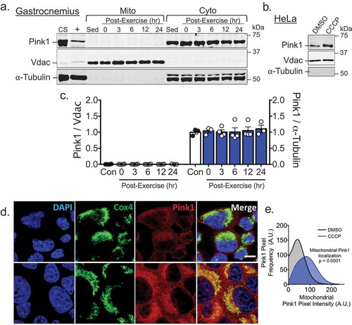
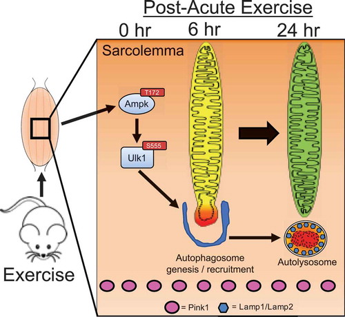
Figure 2. Schematic presentation of timeline for mechanisms regulating acute exercise-induced mitophagy in skeletal muscle.