Figures & data
Figure 1. CCAT1 is upregulated in cervical cancer tissues. (a) The relative expression of CCAT1 in 50 cervical cancer tissues compared with their adjacent normal cervical tissues. N.S., not significant; *P < 0.05, **P < 0.01. (b) The expression of CCAT1 in the cervical cancer tissues compared with the normal cervical tissues. The expression level of CCAT1 was normalized to GAPDH. (c) Statistical analysis of clinical data. SCC, squamous cell carcinoma; AC, adenocarcinoma.
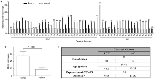
Figure 2. CCAT1 promotes cervical cancer cell proliferation and invasion. (a) The relative expression of CCAT1 in SiHa and HeLa cells with overexpressed or knocked-down CCAT1 compared the controls. (b) Cell viabilities of the SiHa and HeLa cells at 24, 48 and 72 h after overexpressing or knocking down CCAT1. The cell viabilities were determined by the CCK-8 assay. (c) Clone formation images and statistical analysis of the SiHa and HeLa cells transduced with lv-CCAT1 and lv-con. (d) Clone formation images and statistical analysis of the SiHa and HeLa cells transduced with lv-shCCAT1 and lv-shcon. (e) The invasion ability of the SiHa and HeLa cells transduced with lv-CCAT1 and lv-con or (f) transduced with lv-shCCAT1 and lv-shcon. The error bars represent the mean ± SD of the triplicate experiments. *P < 0.05; **P < 0.01.
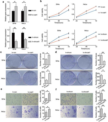
Figure 3. CCAT1-regulated expression of miR-181a-5p and MMP14 in SiHa and HeLa cells. (a) The relative gene expression of miR-181a-5p and MMP14 in the SiHa and HeLa cells transduced with the lv-CCAT1 or (b) lv-shCCAT1 lentivirus and their respective control lentiviruses. (c) Protein expression of MMP14 in the cells transduced with lv-CCAT1 and lv-con. (d) Protein expression of MMP14 in the cells transduced with lv-shCCAT1 and lv-shcon. (e) Protein expression and (f) statistical analysis of MMP14 in cervical cancer tissues and normal cervical tissues. (g) Protein expression of HB-EGF in the cells transduced with lv-CCAT1 and lv-con. (h) Protein expression of HB-EGF in the cells transduced with lv-shCCAT1 and lv-shcon. Lv-CCAT1 and lv-con, CCAT1 overexpression lentivirus and its control lentivirus, respectively. Lv-shCCAT1 and lv-shcon, CCAT1 knockdown lentivirus and its control lentivirus, respectively.
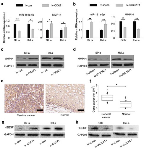
Figure 4. The oncogenic activity of CCAT1 partially negatively regulates miR-181a-5p and then modulates MMP14 and HBEGF. (a) Cell viabilities of the SiHa and HeLa cells transduced with lv-CCAT1 and then transfected with miR-181a-5p mimic or (b) lv-shCCAT1 and then transfected with miR-181a-5p inhibitor. The cell viability was measured by the CCK-8 assay. (c) The relative mRNA expression of CCAT1, MMP14 and HB-EGF in SiHa and HeLa cells transduced with lv-CCAT1 and then transfected with miR-181a-5p mimic or (d) transduced with lv-shCCAT1 and then transfected with miR-181a-5p inhibitor. (e) The protein expression of MMP14 and HB-EGF in SiHa and HeLa cells transduced with lv-CCAT1 and then transfected with miR-181a-5p mimic or (f) transduced with lv-shCCAT1 and then transfected with miR-181a-5p inhibitor. Lv-CCAT1 and lv-con, CCAT1 overexpression lentivirus and its control lentivirus, respectively. Lv-shCCAT1 and lv-shcon, CCAT1 knockdown lentivirus and its control lentivirus, respectively.
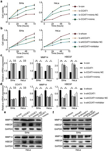
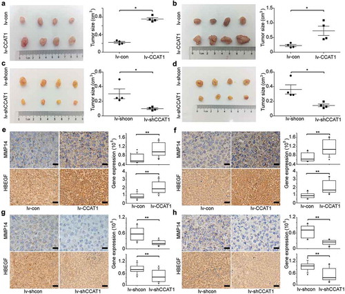
Figure 5. CCAT1 promotes tumor growth of SiHa and HeLa cells. SiHa and HeLa cells were subcutaneously injected in the right flanks of the BALB/c-nu mice. (a) Photographs of the subcutaneously formed tumors and estimated sizes of tumors of SiHa transduced with lv-con/lv-CCAT1 and (c) lv-shcon/lv-shCCAT1. (b) Photographs of the subcutaneously formed tumors and estimated sizes of tumors of HeLa transduced with lv-con/lv-CCAT1 (above) and (d) lv-shcon/lv-shCCAT1. The tumor size of SiHa and HeLa was measured starting 2 weeks after injection. (e) Representative images of IHC staining and a comparison of the protein expression of MMP14 and HB-EGF in xenografts of SiHa and (f) HeLa transduced with lv-con/lv-CCAT1. (g) Representative images of IHC staining and a comparison of the protein expression of MMP14 and HB-EGF in xenografts of SiHa and (h) HeLa transduced with lv-shcon/lv-shCCAT1.