Figures & data
Figure 1. Effect of sera from the Jixuepaidu Tang-1-treated mice on the viability, apoptosis, and EMT process in HG-treated podocytes
The podocytes were cultured with media containing normal glucose (NG; 5.6 mmol/L D-glucose), high glucose (HG; 30 mmol/L D-glucose), sera from the control physiological saline-treated mice (CS), and sera from the low/mid/high dose of Jixuepaidu Tang-1-treated mice (LD/MD/HD). With the exception of the NG group, the podocytes in the other groups were stimulated with HG. Following 24 h of culture, (a) the cell viability was determined using the MTT method. (b) The cell apoptosis was determined using the TUNEL staining. The apoptosis index (AI) was evaluated as the percentage of TUNEL-positive nuclei (brown) in sections. (c) Representative western blot analysis of nephrin, P-cadherin, desmin, and vimentin expression in podocytes. The graph represents the densitometric analysis normalized to β-actin. (d) Representative immunofluorescence images of the slit diaphragm protein nephrin (green) and podocyte injury marker desmin (red). Cell nucleus was labeled in blue by DAPI. Scale bar: 25 μm. Data are presented as mean ± SD; n = 3; *p < 0.05 vs. NG; #p < 0.05 vs. CS.
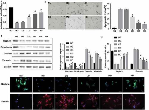
Figure 2. Jixuepaidu Tang-1 sera inhibited the HG-induced podocyte injury and EMT through blocking the TGF-β1/SGK1 signaling
(a) Representative western blot analysis of TGF-β1 and SGK1 expression in podocytes in the groups treated the same as in . The graph represents the densitometric analysis normalized to β-actin. *p < 0.05 vs. NG; #p < 0.05 vs. CS. (b–e) The podocytes were cultured with media containing normal glucose (NG; 5.6 mmol/L D-glucose), high glucose (HG; 30 mmol/L D-glucose), sera from the high dose of Jixuepaidu Tang-1-treated mice (HD), TGF-β1 recombinant protein (TGF-β1), or SGK1 recombinant protein (SGK1) (Thermo Scientific). The podocytes in the si-Ctrl and si-SGK1 groups were transfected with si-Ctrl and si-SGK1 respectively before treatment with HG and HD. With the exception of the NG group, the podocytes in the other groups were stimulated with HG. Following 24 h of culture, (b) the cell viability was determined using the MTT method. (c) The cell apoptosis was determined using the TUNEL staining. The apoptosis index (AI) was evaluated as the percentage of TUNEL-positive nuclei (brown) in sections. (d) Representative western blot analysis of nephrin, P-cadherin, desmin, vimentin, TGF-β1, and SGK1 expression in podocytes. Graph represents the densitometric analysis normalized to β-actin. (e) Representative immunofluorescence images of nephrin (green) and desmin (red). Cell nucleus was labeled in blue by DAPI. Scale bar: 25 μm. Data are presented as mean ± SD; n = 3; (B-E) *p < 0.05 vs. NG; #p < 0.05 vs. HG; &p < 0.05 vs. HD; $p < 0.05 vs. HD+si-Ctrl.
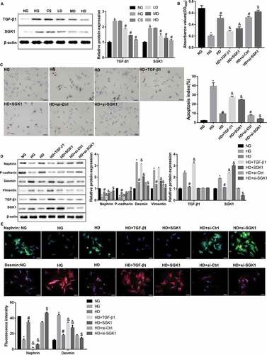
Figure 3. LOC498759 was involved in the Jixuepaidu Tang-1 sera-mediated inhibition of the HG-induced podocyte injury and EMT
qRT-PCR was performed to detect relative LOC498759 expression in podocytes in the groups treated the same as in . *p < 0.05 vs. NG; #p < 0.05 vs. HG; &p < 0.05 vs. HD; $p < 0.05 vs. HD+si-Ctrl. (b) Treatment with TGF-β1 and SGK1 recombinant proteins significantly increased LOC498759 expression. *p < 0.05 vs. Control. (c–g) The podocytes were transfected with LOC498759 overexpression vector and control vector, or si-LOC498759 and scramble si-Ctrl, followed by treatment with high glucose (HG; 30 mmol/L D-glucose) and sera from the high dose of Jixuepaidu Tang-1-treated mice (HD). (c) qRT-PCR was performed to examine the knockdown or overexpression efficiency. Following 24 h of culture, (d) the cell viability was determined using the MTT method. (e) The cell apoptosis was determined using the TUNEL staining. The apoptosis index (AI) was evaluated as the percentage of TUNEL-positive nuclei (brown) in sections. (f) Representative western blot analysis of nephrin, P-cadherin, desmin, and vimentin, expression in podocytes. The graph represents the densitometric analysis normalized to β-actin. (G) Representative immunofluorescence images of nephrin (green) and desmin (red). Cell nucleus was labeled in blue by DAPI. Scale bar: 25 μm. Data are presented as mean ± SD; n = 3; (C-G) *p < 0.05 vs. HG+HD+vector; #p < 0.05 vs. HG+HD+si-Ctrl.
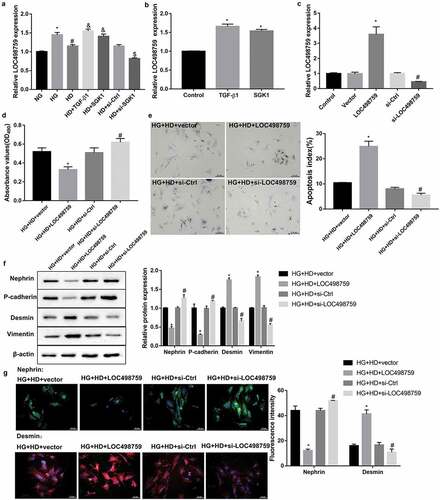
Table 1. Metabolic data of mice at the end of the experiment (mean ± SD)
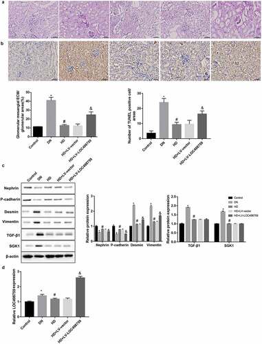
Figure 4. LOC498759 was involved in the Jixuepaidu Tang-1-mediated renal protection in DN mice
C57BL/6J mice were randomly divided into 5 groups: normal control group (Control), DN model group (DN), high dose of Jixuepaidu Tang-1 group (HD), high dose of Jixuepaidu Tang-1 + empty lentivirus group (HD + LV-vector) group, and high dose of Jixuepaidu Tang-1 + LOC498759 lentivirus group (HD + LV-LOC498759). At the end of the experiment, the mice were sacrificed and the kidneys were separated for the following analysis. (a) The glomerular mesangial extracellular matrix (ECM) deposition in mouse kidney tissues was assessed using PAS staining. The percentage of glomerular mesangial ECM/glomerular area was calculated. Scale bar: 25 μm. (b) The apoptosis of glomerular cells in mouse kidney tissues was examined using TUNEL staining. The number of TUNEL positive cells (brown) was counted. Scale bar: 25 μm. (c) Representative western blot analysis of nephrin, P-cadherin, desmin, vimentin, TGF-β1, and SGK1 expression in mouse kidney tissues. The graph represents the densitometric analysis normalized to β-actin. (d) qRT-PCR was performed to detect relative LOC498759 expression in mouse kidney tissues. *p < 0.05 vs. control; #P < 0.05 vs. DN; &p < 0.05 vs. LV-vector.