Figures & data
Figure 1. miR-34b/c and ITCH are required for Dox-induced myocardial injury. (a-b). HL-1 cells treated by Dox at the indicated concentrations (0, 0.01, 0.1, 1 and 10 μM) were observed under an inverted microscope, and HL-1 cell viability was gradually reduced after the treatment of Dox at the indicated concentrations. (c). mRNA expression of ITCH was gradually down-regulated after the treatment of Dox at the indicated concentrations. (d-e). mRNA expressions of TNF-α and IL-6 were gradually up-regulated after the treatment of Dox at the indicated concentrations. (f-g). miR-34b and miR-34c expressions were gradually up-regulated after the treatment of Dox at the indicated concentrations. (h). miR-34b expression had positive correlation with the expression of miR-34c. (i). miR-34b expression had negative correlation with the mRNA expression of ITCH. (j). miR-34c expression had negative correlation with the mRNA expression of ITCH. *P < 0.05 vs control; **P < 0.01 vs control
Figure 2. Doxorubicin activates NF-κB pathway in HL-1 cells. HL-1 cells were treated by Dox (0.05 μM). (a). MTT assay showed that HL-1 cell viability was reduced after the treatment of Dox. (b). TUNEL staining showed that TUNEL positive rate was 44.2%. (c). Protein level of ITCH was reduced in HL-1 cells treated by Dox, whereas protein level of NF-κB was increased in nucleus or cytoplasm of HL-1 cells treated by Dox. (d). Under the treatment of Dox, BAY 11–7082 (5 μM) was added to HL-1 cells and cultured for 20 h. TNF-α and IL-6 levels increased in the supernatant of HL-1 cells. (e). Silencing ITCH in HL-1 cells promoted Dox-induced total NF-κB and pNF-κB protein levels. (f). HL-1 cells were treated by Dox for 0, 4, 8, 12 and 20 h. Silencing ITCH in HL-1 cells promoted protein levels of NF-κB, TNF-α and IL-6. **P < 0.01 vs control
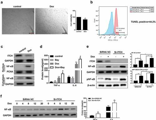
Figure 3. The regulation of miR-34b/c on ITCH. (a-b). There were binding sites between miR-34b/c and ITCH. Dual-luciferase reporter assay showed that miR-34b/c mimic significantly decreased the luciferase activity of pmirGLO-ITCH, whereas miR-34b/c mimic did not significantly change the luciferase activity of pmirGLO-mut-ITCH. **P < 0.01 vs pmirGLO-ITCH+miRNC. (c-h). Under the treatment of Dox, miR-34b/c mimic reduced protein level of ITCH, promoted nucleus and cytoplasm protein level of NF-κB, increased protein and mRNA expression of TNF-α and IL-6, and decreased HL-1 cell viability. **P < 0.01 vs Dox; ##P < 0.01 vs miR-34b/c mimic
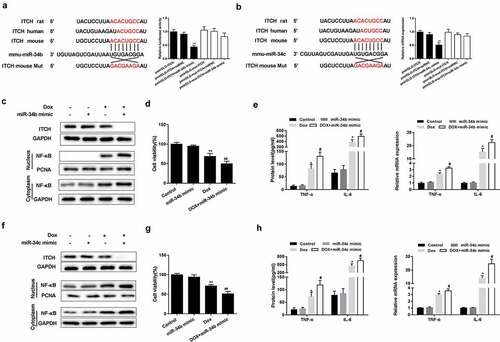
Figure 4. Silencing miR-34 protected HL-1 cells through regulating ITCH. (a). Under the treatment of Dox, miR-34b inhibitor reversed the Dox-induced decrease of ITCH protein level and promotion of NF-κB pathway, whereas si-ITCH could not reverse the effects induced by Dox. (b). miR-34b inhibitor reversed the Dox-induced increase of mRNA expression of TNF-α and IL-6, whereas si-ITCH could not reverse the effects induced by Dox. (c). miR-34c inhibitor reversed the Dox-induced decrease of ITCH protein level and promotion of NF-κB pathway, whereas si-ITCH could not reverse the effects induced by Dox. (d). miR-34c inhibitor reversed the Dox-induced increase of mRNA expression of TNF-α and IL-6, whereas si-ITCH could not reverse the effects induced by Dox. **P < 0.01 vs Dox; ##P < 0.01 vs Dox+miR-34b/c inhibitor
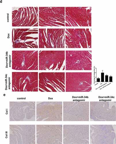
Figure 5. miR-34 Antagomir protected myocardial cells in mouse model of cardiomyopathy. Mice were divided into control (n = 5), Dox-induced cardiomyopathy group (n = 6), Dox+miR-34b antagomir group (n = 6) and Dox+miR-34c antagomir group (n = 6). (a). In Dox group, TNF-α, IL-6, c-caspase3 and NF-κB protein levels were higher in heart tissue, and ITCH protein level was lower in heart tissue. miR-34b/c antagomir treatment could reverse these effects. (b). Immunohistochemistry showed that TNF-α and IL-6 levels were higher in heart tissue, miR-34b/c antagomir treatment decreased TNF-α and IL-6 levels. (c). HE staining showed that in Dox group, myofibrillar degeneration and disruption was observed. More normal morphology and myofibrosis were observed in Dox+miR-34b/c antagomir groups than Dox group, and myofibrillar degeneration and disruption were relieved in Dox+miR-34b/c antagomir groups than Dox group. (d). MASSON staining showed that myofibrosis was induced in the Dox group, and miR-34b/c antagomir treatment relieved myofibrosis. E. Collagen type 1 (col I) and collagen type 3 (col III) expressions were detected by Immunohistochemistry. *P < 0.05 vs control
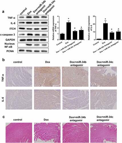
Figure 5. (continued)