
Figures & data
Table 1. Sequences used in cell transfection
Figure 1. Circ_0001649 was down-regulated in OS. Circ_0001649 expression was measured through qRT-PCR in OS tissues (a) and cell lines (b). * P < 0.05, ** P < 0.01 and *** P < 0.001 contrasted with indicated group or hFOB1.19 group
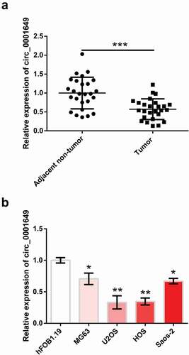
Figure 2. Circ_0001649 overexpression inhibited the growth of U2OS and HOS cells, which were transfected with pEX-circ_0001649. (a) Expression of circ_0001649 was detected by qRT-PCR. Viability (b) and survival fraction (c) were detected by CCK-8 and colony formation assay. (d–e) Levels of CDK4 and CDK6 were detected by western blot analysis. (f) Apoptosis was detected by flow cytometry. (g–h) Levels of apoptosis-related factors were detected by western blot analysis. * P < 0.05, ** P < 0.01 and *** P < 0.001 contrasted with indicated group
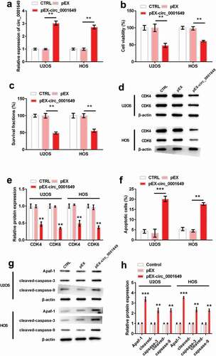
Figure 3. Circ_0001649 could sponge multiple miRNAs. (a) MiRNA expression in si-circ_0001649 transfected U2OS cell was detected by qRT-PCR. Luciferase activity experiment detected target relationship between circ_0001649 and miR-338-5p (b), miR-647 (c) and miR-942 (d), respectively. (e) The potential sequences of circ_0001649 and miR-338-5p, miR-647 and miR-942, respectively. * P < 0.05, ** P < 0.01 and *** P < 0.001 contrasted with indicated group
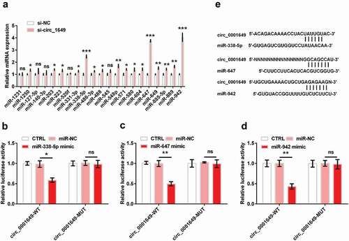
Figure 4. Circ_0001649 inhibited U2OS and HOS cells proliferation through sponging miR-338-5p, miR-647 and miR-942 after cells were co-transfected with pEX-circ_0001649 and miRNAs mimic. Cell viability was detected through CCK-8 in U2OS (a) and HOS (b) cells. Survival fraction was tested through colony formation in U2OS (c) and HOS (d) cells. Expression of CDK4 and CDK6 was detected through western blot and western blot quantitation in U2OS (e, f) and HOS (g, h) cells. * P < 0.05 and ** P < 0.01 contrasted with indicated group
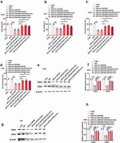
Figure 5. Circ_0001649 promoted U2OS and HOS cells apoptosis through sponging miR-338-5p, miR-647 and miR-942 after cells were co-transfected with pEX-circ_0001649 and miRNAs mimic. Apoptosis was detected through flow cytometry in U2OS (a) and HOS (b) cells. Expression of apoptosis-related factors was detected through western blot and western blot quantitation in U2OS (c, d) and HOS (e, f) cells. * P < 0.05, ** P < 0.01 and *** P < 0.001 contrasted with indicated group
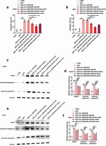
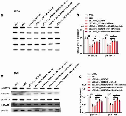
Figure 6. The inhibition of STAT signal pathway by circ_0001649 was detected after co-transfected with pEX-circ_0001649 and miRNAs mimic in U2OS and HOS cells. Expression of STAT3 and STAT5 was detected through western blot analysis (a, b) in U2OS cell. Expression of STAT3 and STAT5 was detected through western blot analysis (c, d) in HOS cell. * P < 0.05, ** P < 0.01 and *** P < 0.001 contrasted with indicated group
Data availability statement
The datasets used and/or analyzed during the current study are available from the corresponding author on reasonable request.