
Figures & data
Figure 1. Autophagy was elevated in gastric cancer stem cells (GCSCs)
(a) GCSCs were isolated from gastric cancer cell lines (BGC-823 and SGC-7901) using an anti‐CD44 antibody. The expression levels of (b) miR-200b, (c) protein kinase C α (PKCα), and (d) RAB37 were determined in CD44− and CD44+ BGC-823 and SGC-7901 cells by qRT-PCR. The protein levels of (e) PKCα and RAB37; (f) microtubule-associated protein light chain 3 (LC3) I, LC3II, and P62 were determined in CD44− and CD44+ BGC-823 and SGC-7901 cells by western blot, GAPDH was used as an internal control. The LC3II/LC3I ratio was calculated by Image J.*P < 0.05 vs CD44−.

Figure 2. MET increased the methylation and phosphorylation of RAB37 and suppressed autophagy in GCSCs
The CD44+ BGC-823 and CD44+SGC-7901 (hereinafter referred to as BGC-823 and SGC-7901) were cultured in MET−homocysteine (HCY)+ or MET+HCY− medium. The expressions of (a) PKCα, (b) miR-200b, and (c) RAB37 were determined by qRT-PCR. (d) The cell viability was assessed by methyl thiazolyl tetrazolium (MTT) assay. (e) The methylation levels of RAB37 and miR-200b promoters were measured by Methylation-specific RCR (MSP). U results with primers specific for unmethylated sequences.M results with primers specific for methylated sequences. The protein levels of (f) PKCα, P-RAB37 and RAB37; (g) LC3I, LC3II, and P62 were determined by western blot, GAPDH was used as an internal control. The LC3II/LC3I ratio was calculated by Image J. (h and i) The cell apoptosis was assessed by flow cytometry.*P < 0.05 vs MET−HCY+.
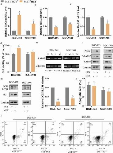
Figure 3. Metase supplementation reduced the methylation and phosphorylation of RAB37 and promoted autophagy in GCSCs
BGC-823 and SGC-7901 cells were cultured in MET− HCY+ or MET+HCY− medium with or without the infections of lentivirus vector (LV)-Metase or its negative control (LV-control). The expressions of (a) RAB37, (b) miR-200b, and (c) PKCα were determined by qRT-PCR. (d) The cell viability was assessed by MTT assay. (e) The methylation levels of RAB37 and miR-200b promoters were measured by MSP. U results with primers specific for unmethylated sequences. M results with primers specific for methylated sequences. The protein levels of (f) PKCα, P-RAB37 and RAB37; (g) LC3I, LC3II, and P62 were determined by western blot, GAPDH was used as an internal control. The LC3II/LC3I ratio was calculated by Image J.*P < 0.05 vs MET−HCY+, #P < 0.05 vs MET+HCY−+LV-control.
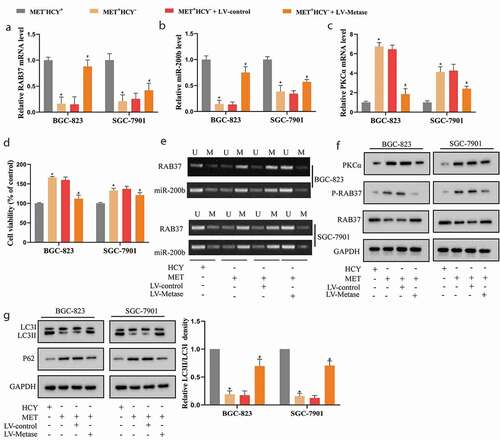
Figure 4. miR-200b negatively regulated PKCα expression in GCSCs
(a) An online bioinformatics database (http://starbase.sysu.edu.cn/) predicted the binding sites between miR-200b and PKCα. (b) The luciferase activities of wild type PKCα (wt) and mutation PKCα (mut) were detected in 293 T cells which were transfected with miR-200b mimic or its negative control (NC mimic). The expressions of (c) miR-200b and (d) PKCα in BGC-823 and SGC-7901 cells which were transfected with miR-200b mimic or miR-200b inhibitor were measured by qRT-PCR. (e) The protein level of PKCα in BGC-823 and SGC-7901 cells which were transfected with miR-200b mimic or miR-200b inhibitor was measured by western blot, GAPDH was used as an internal control.*P < 0.05 vs NC mimic, #P < 0.05 vs NC inhibitor.
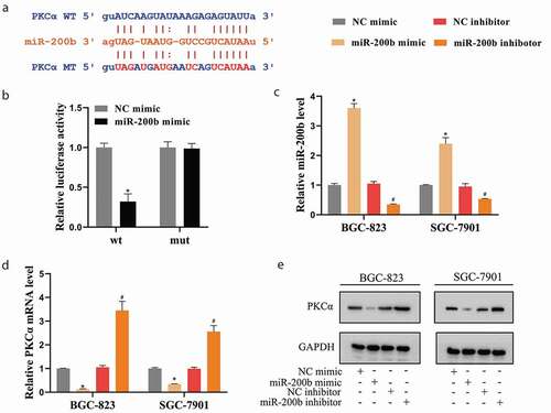
Figure 5. miR-200b/PKCα axis was involved in the promoting effect of Metase on autophagy of GCSCs by regulating the phosphorylation level of RAB37
The BGC-823 and SGC-7901 cells were transfected with LV-Metase+miR-200b inhibitor or LV-Metase+miR-200b inhibitor+si-PKCα or LV-Metase+corresponding controls (NC inhibitor or si-RNA) and then all cells were cultured in MET+HCY− medium. The expressions of (a) RAB37, (b) PKCα, and (c) miR-200b were determined by qRT-PCR. (d) The cell viability was assessed by MTT assay. The protein levels of (e) PKCα, P-RAB37 and RAB37; (f) LC3I, LC3II, and P62 were determined by western blot, GAPDH was used as an internal control. The LC3II/LC3I ratio was calculated by Image J.*P < 0.05 vs MET+HCY−+ LV-Metase+NC inhibitor, #P < 0.05 vs MET+HCY−+LV-Metase+ miR-200b inhibitor+si-RNA.
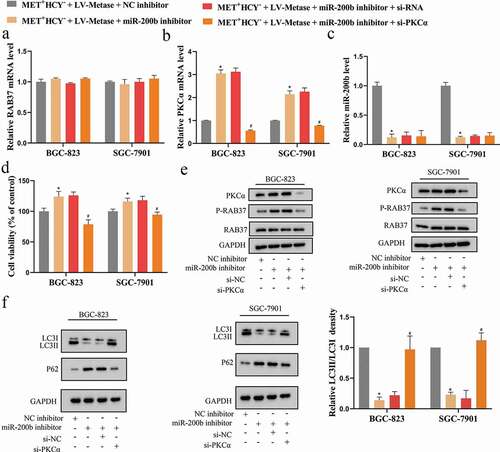
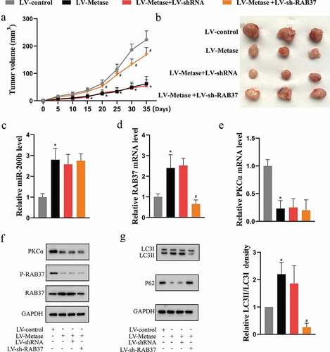
Figure 6. Metase suppressed tumor growth by enhancing the expression and activity of RAB37 in vivo.
The xenograft mice were randomly divided into 4 groups: LV-control (n = 8), LV-Metase (n = 8), LV-Metase+LV-sh-RAB37 (n = 8), and LV-Metase+LV-sh-RNA (n = 8). (a) The tumor volume of each mice was measured. (b) The representative tumor pictures of mice in each group. The expressions of (c) miR-200b, (d) RAB37, and (e) PKCα were determined by qRT-PCR. The protein levels of (f) PKCα, P-RAB37 and RAB37; (g) LC3I, LC3II, and P62 were determined by western blot, GADPH was used as an internal control. *P < 0.05 vs LV-control, #P < 0.05 vs LV-Metase+LV-sh-RNA.
Supplemental material