Figures & data
Figure 1. LPS-induced TRIM31 upregulation and cell apoptosis. (a) Real-time PCR was performed to measure the relative mRNA expression of TRIM31 in peripheral blood samples from 35 septic patients and 25 normal patients. *, P < 0.05, ***, P < 0.001 vs normal persons. (b) Cell proliferation was analyzed after stimulation with LPS (three repetitions). (c) Cell apoptosis was analyzed after LPS treatment. (d, e): The mRNA and protein levels of TRIM31 were measured by qRT-PCR (d) and Western blot (e). ***, P < 0.001 vs 0 μg/ml LPS
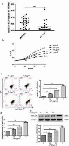
Figure 2. TRIM31 knockdown inhibited cell apoptosis induced by LPS. (a, b): The mRNA and protein levels of TRIM31 were measured by qRT-PCR (a) and Western blot (b). (c): Cell apoptosis was measured by flow cytometry. (d): ELISA was used to measure the concentrations of IL-1β and TNF-α. (e): The protein levels of NF-κB signaling related protein were measured by Western blot
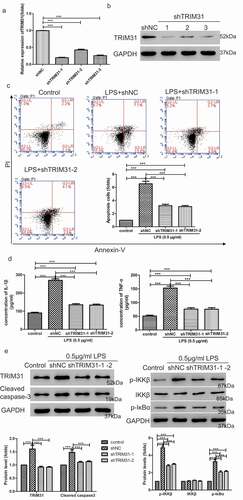
Figure 3. TRIM31 elevated cell apoptosis induced by LPS. (a, b): AC16 cells were transduced with TRIM31 overexpression or vector lentivirus. Overexpression efficiency was analyzed by qRT-PCR (a) and Western blot (b). (c): Annexin V/PI assay was used to assess cell apoptosis. (d): ELISA was used to measure the concentrations of IL-1β and TNF-α. (e): The protein levels of NF-κB signaling related protein were measured by Western blot
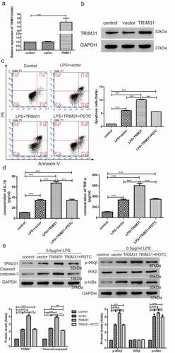
Figure 4. TAK1-mediated activation of NF-κB signaling pathway induced by TRIM31. (a): Western blot was used to determine the protein levels of TAK1 and p-TAK1. (b): Cell apoptosis was measured by flow cytometry. (c): The protein levels of cleaved-caspase-3, p-IKKβ and p-IκBα were measured by Western blot. (d): ELISA was used to measure the concentrations of IL-1β and TNF-α. (e): Co-immunoprecipitation assay was used to examine the interaction between TRIM31 and TAK1. (f): The ubiquitin level of TAK1 was measured
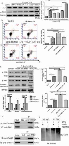
Figure 5. TRIM31 deletion suppressed apoptosis in mice model of sepsis induced by LPS. (a) H&E staining was performed on heart tissue samples of animal models. (b) IHC was used to detect the protein level of NF-κB (c) TUNEL assay was performed to measure apoptosis in heart tissue samples of animal models. a, control; b, LPS + vector; c, LPS + shTRIM31; d, LPS + PDTC
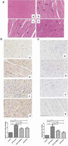
Figure 6. IHC was used to detect the protein levels of p-IKKβ (a) and p-IκBα (b). a, control; b, LPS + vector; c, LPS + shTRIM31; d, LPS + PDTC
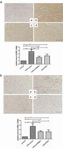
Figure 7. TRIM31 deletion blocked LPS-induced activation of NF-κB signaling pathway. (a): ELISA was used to measure the concentrations of IL-1β and TNF-α. (b) The protein levels of NF-κB signaling related protein were measured by Western blot